Login
Registration enables users to use special features of this website, such as past
order histories, retained contact details for faster checkout, review submissions, and special promotions.
order histories, retained contact details for faster checkout, review submissions, and special promotions.
Forgot password?
Registration enables users to use special features of this website, such as past
order histories, retained contact details for faster checkout, review submissions, and special promotions.
order histories, retained contact details for faster checkout, review submissions, and special promotions.
Quick Order
Products
Antibodies
ELISA and Assay Kits
Research Areas
Infectious Disease
Resources
Purchasing
Reference Material
Contact Us
Locations
Orders Processing,
Shipping & Receiving,
Warehouse
2 Shaker Rd Suites
B001/B101
Shirley, MA 01464
Production Lab
Floor 6, Suite 620
20700 44th Avenue W
Lynnwood, WA 98036
Telephone Numbers
Tel: +1 (206) 374-1102
Fax: +1 (206) 577-4565
Contact Us
Additional Contact Details
Login
Registration enables users to use special features of this website, such as past
order histories, retained contact details for faster checkout, review submissions, and special promotions.
order histories, retained contact details for faster checkout, review submissions, and special promotions.
Forgot password?
Registration enables users to use special features of this website, such as past
order histories, retained contact details for faster checkout, review submissions, and special promotions.
order histories, retained contact details for faster checkout, review submissions, and special promotions.
Quick Order
| Catalog Number | Size | Price |
|---|---|---|
| LS-C745593-100 | 100 µg (1 mg/ml) | $310 |
1 of 3
2 of 3
3 of 3
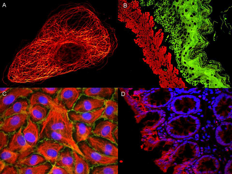
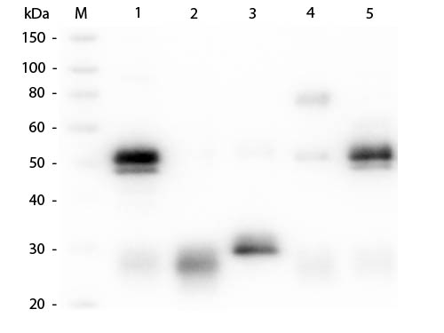
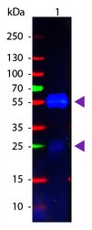
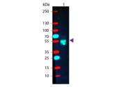
Polyclonal Goat Anti‑Rabbit IgG Secondary Antibody (Atto 488, IF, WB) LS‑C745593
Polyclonal Goat Anti‑Rabbit IgG Secondary Antibody (Atto 488, IF, WB) LS‑C745593
Antibody:
Rabbit IgG Goat anti-Rabbit Polyclonal (Atto 488) Antibody
Application:
IF, WB, FLISA
Reactivity:
Rabbit
Format:
Atto 488, Unmodified
Toll Free North America
 206-374-1102
For Research Use Only
Overview
Antibody:
Rabbit IgG Goat anti-Rabbit Polyclonal (Atto 488) Antibody
Application:
IF, WB, FLISA
Reactivity:
Rabbit
Format:
Atto 488, Unmodified
Specifications
Description
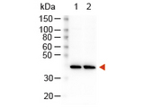
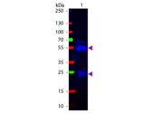
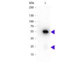
Rabbit IgG antibody LS-C745593 is an Atto 488-conjugated goat polyclonal antibody. Validated for FLISA, IF and WB.
Host
Goat
Reactivity
Rabbit
(tested or 100% immunogen sequence identity)
Clonality
IgG
Polyclonal
Conjugations
Atto 488
Purification
Affinity chromatography
Modifications
Unmodified
Immunogen
Rabbit IgG whole molecule
Specificity
Assay by immunoelectrophoresis resulted in a single precipitin arc against anti-Goat Serum, Rabbit IgG and Rabbit Serum. No reaction was observed against Bovine, Chicken, Goat, Guinea Pig, Hamster, Horse, Human, Mouse, Rat and Sheep Serum Proteins. This antibody will react with heavy chains of rabbit IgG and with light chains of most rabbit immunoglobulins.
Applications
- Immunofluorescence (1:5000)
- Western blot (1:10000)
- Fluorophore-Linked Immunosorbent Assay (1:20000)
Usage
Applications should be user optimized.
Presentation
Lyophilized from 10 mg/mL BSA (Protease and Ig free), 0.02 M Potassium Phosphate, pH 7.2, 0.15 M NaCl, 0.01% Sodium Azide
Reconstitution
Reconstitute in 100 µL of deionized water.
Storage
Store vial at 4°C prior to restoration. For extended storage aliquot contents and freeze at -20°C or below. Avoid freeze-thaw cycles. Stable for several weeks at 4°C as an undiluted liquid. Dilute only prior to immediate use.
Restrictions
For research use only. Intended for use by laboratory professionals.
Publications (0)
Customer Reviews (0)
Featured Products
Species:
Rabbit
Applications:
Immunofluorescence, Western blot, Flow Cytometry
Species:
Rabbit
Applications:
IHC, Western blot, ELISA
Species:
Rabbit
Applications:
Immunofluorescence, Western blot, Flow Cytometry
Species:
Rabbit
Applications:
IHC, Western blot, ELISA, Dot Blot, Electron Microscopy
Species:
Rabbit
Applications:
IHC - Frozen, Western blot, ELISA
Species:
Rabbit
Applications:
IHC, Western blot, ELISA
Request SDS/MSDS
To request an SDS/MSDS form for this product, please contact our Technical Support department at:
Technical.Support@LSBio.com
Requested From: United States
Date Requested: 4/17/2024
Date Requested: 4/17/2024


