Login
Registration enables users to use special features of this website, such as past
order histories, retained contact details for faster checkout, review submissions, and special promotions.
order histories, retained contact details for faster checkout, review submissions, and special promotions.
Forgot password?
Registration enables users to use special features of this website, such as past
order histories, retained contact details for faster checkout, review submissions, and special promotions.
order histories, retained contact details for faster checkout, review submissions, and special promotions.
Quick Order
Products
Antibodies
ELISA and Assay Kits
Research Areas
Infectious Disease
Resources
Purchasing
Reference Material
Contact Us
Locations
Orders Processing,
Shipping & Receiving,
Warehouse
2 Shaker Rd Suites
B001/B101
Shirley, MA 01464
Production Lab
Floor 6, Suite 620
20700 44th Avenue W
Lynnwood, WA 98036
Telephone Numbers
Tel: +1 (206) 374-1102
Fax: +1 (206) 577-4565
Contact Us
Additional Contact Details
Login
Registration enables users to use special features of this website, such as past
order histories, retained contact details for faster checkout, review submissions, and special promotions.
order histories, retained contact details for faster checkout, review submissions, and special promotions.
Forgot password?
Registration enables users to use special features of this website, such as past
order histories, retained contact details for faster checkout, review submissions, and special promotions.
order histories, retained contact details for faster checkout, review submissions, and special promotions.
Quick Order
| Catalog Number | Size | Price |
|---|---|---|
| LS-C154186-1 | 1 mg | $768 |
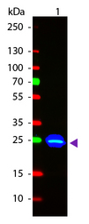
![GFP Antibody - Anti-GFP Antibody - Western Blot. Western blot of GFP recombinant protein detected with polyclonal anti-GFP antibody. Lane 1 shows detection of a 33 kD band corresponding to a GFP containing recombinant protein (arrowhead) expressed in HeLa cells. Lane 2 shows no staining of a mock transfected HeLa cell lysate. A 4-12% Bis-Tris gradient gel was used for SDS-PAGE. The protein was transferred to nitrocellulose using standard methods. After blocking the membrane was probed with the primary antibody diluted to 1 ug/ml for 1 h at room temperature followed by washes and reaction with a 1:2500 dilution of IRDye 800 conjugated Donkey-a-Goat IgG [H&L] MX7 (. The IRDye 800 fluorescence image was captured using the Odyssey Infrared Imaging System developed by LI-COR. IRDye is a trademark of LI-COR, Inc. Other detection systems will yield similar results. This image was taken for the unconjugated form of this product. Other forms have not been tested.](https://lsbio-7d62.kxcdn.com/image2/gfp-antibody-fitc-ls-c154186/81887_126461.jpg)
1 of 2
2 of 2
Polyclonal Goat anti‑Aequorea victoria GFP Antibody (FITC, IF, WB) LS‑C154186
Polyclonal Goat anti‑Aequorea victoria GFP Antibody (FITC, IF, WB) LS‑C154186
Antibody:
GFP Goat anti-Aequorea victoria Polyclonal (FITC) Antibody
Application:
IF, WB, FLISA
Reactivity:
Aequorea victoria
Format:
FITC, Unmodified
Toll Free North America
 206-374-1102
For Research Use Only
Overview
Antibody:
GFP Goat anti-Aequorea victoria Polyclonal (FITC) Antibody
Application:
IF, WB, FLISA
Reactivity:
Aequorea victoria
Format:
FITC, Unmodified
Specifications
Description
GFP antibody LS-C154186 is an FITC-conjugated goat polyclonal antibody to aequorea victoria GFP. Validated for FLISA, IF and WB.
Host
Goat
Reactivity
Aequorea victoria
(tested or 100% immunogen sequence identity)
Clonality
IgG
Polyclonal
Conjugations
FITC
Purification
Affinity chromatography
Modifications
Unmodified
Immunogen
Recombinant Green Fluorescent Protein (GFP) fusion protein corresponding to the full length amino acid sequence (246aa) derived from the jellyfish Aequorea victoria.
Specificity
Assay by immunoelectrophoresis resulted in a single precipitin arc against anti-Goat Serum, anti-Fluorescein and purified and partially purified Green Fluorescent Protein (Aequorea victoria). No reaction was observed against Human, Mouse and Rat Serum Proteins.
Applications
- Immunofluorescence (1:500 - 1:2500)
- Western blot (1:10000)
- Fluorophore-Linked Immunosorbent Assay (1:20000)
Usage
The applications listed have been tested for the unconjugated form of this product. Other forms have not been tested.
Presentation
Lyophilized from 10 mg/mL BSA (Protease and Ig free), 0.02 M Potassium Phosphate, pH 7.2, 0.15 M NaCl, 0.01% Sodium Azide
Reconstitution
Reconstitute in 1 mL of deionized water.
Storage
Store lyophilized at 4°C. Once reconstituted, aliquot and store at -20°C. Avoid freeze-thaw cycles. Store undiluted. Protect from light.
Restrictions
For research use only. Intended for use by laboratory professionals.
Publications (0)
Customer Reviews (0)
Featured Products
Species:
Aequorea victoria
Applications:
IHC - Paraffin, IHC - Frozen, Western blot, ELISA
Species:
Aequorea victoria
Applications:
IHC, IHC - Paraffin, Western blot, Immunoprecipitation, ELISA
Species:
Aequorea victoria
Applications:
IHC, IHC - Paraffin, IHC - Frozen, Western blot, ELISA
Species:
Aequorea victoria
Applications:
IHC, Immunofluorescence, Western blot, ELISA

Species:
Aequorea victoria
Applications:
IHC, Immunofluorescence, Western blot, Flow Cytometry, ELISA
Request SDS/MSDS
To request an SDS/MSDS form for this product, please contact our Technical Support department at:
Technical.Support@LSBio.com
Requested From: United States
Date Requested: 4/19/2024
Date Requested: 4/19/2024


