Login
Registration enables users to use special features of this website, such as past
order histories, retained contact details for faster checkout, review submissions, and special promotions.
order histories, retained contact details for faster checkout, review submissions, and special promotions.
Forgot password?
Registration enables users to use special features of this website, such as past
order histories, retained contact details for faster checkout, review submissions, and special promotions.
order histories, retained contact details for faster checkout, review submissions, and special promotions.
Quick Order
Products
Antibodies
ELISA and Assay Kits
Research Areas
Infectious Disease
Resources
Purchasing
Reference Material
Contact Us
Locations
Orders Processing,
Shipping & Receiving,
Warehouse
2 Shaker Rd Suites
B001/B101
Shirley, MA 01464
Production Lab
Floor 6, Suite 620
20700 44th Avenue W
Lynnwood, WA 98036
Telephone Numbers
Tel: +1 (206) 374-1102
Fax: +1 (206) 577-4565
Contact Us
Additional Contact Details
Login
Registration enables users to use special features of this website, such as past
order histories, retained contact details for faster checkout, review submissions, and special promotions.
order histories, retained contact details for faster checkout, review submissions, and special promotions.
Forgot password?
Registration enables users to use special features of this website, such as past
order histories, retained contact details for faster checkout, review submissions, and special promotions.
order histories, retained contact details for faster checkout, review submissions, and special promotions.
Quick Order
| Catalog Number | Size | Price |
|---|---|---|
| LS-C154208-1 | 1 mg (1 mg/ml) | $921 |
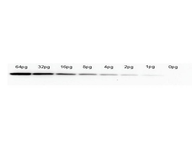
![GFP Antibody - Anti-GFP Antibody - Western Blot. Western blot of GFP recombinant protein detected with monoclonal anti-GFP antibody. GFP recombinant protein was expressed in HeLa cells, where 50 ng (lane 1), 100 ng (lane 2) and 500 ng (lane 3) of lysate were loaded per lane. Mab anti-GFP detects a 27 kD band corresponding to the epitope tag GFP. The cell lysates were prepared in a RIPA buffer containing 200 mM NaCl. A 4-12% Bis-Tris gradient gel (Invitrogen) was used for SDS-PAGE. The protein was transferred to nitrocellulose using standard methods. After blocking with 5% BLOTTO in PBS, the membrane was probed with the primary antibody diluted to 1.0 mg/ml for 1 h at room temperature followed by washes and reaction with a 1:2500 dilution of IRDye 800 conjugated Goat-a-Mouse IgG [H&L] MX10 (. IRDye 800 fluorescence image was captured using the Odyssey Infrared Imaging System developed by LI-COR. IRDye is a trademark of LI-COR, Inc. Other detection systems will yield similar results.](https://lsbio-7d62.kxcdn.com/image2/gfp-antibody-clone-9f9.f9-ls-c154208/81891_126465.jpg)
1 of 2
2 of 2
Monoclonal Mouse anti‑Aequorea victoria GFP Antibody (clone 9F9.F9, IHC, IF, WB) LS‑C154208
Monoclonal Mouse anti‑Aequorea victoria GFP Antibody (clone 9F9.F9, IHC, IF, WB) LS‑C154208

Antibody:
GFP Mouse anti-Aequorea victoria Monoclonal (9F9.F9) Antibody
Application:
IHC, IF, WB, Flo, ELISA
Reactivity:
Aequorea victoria
Format:
Unconjugated, Unmodified
Toll Free North America
 206-374-1102
For Research Use Only
Overview
Antibody:
GFP Mouse anti-Aequorea victoria Monoclonal (9F9.F9) Antibody
Application:
IHC, IF, WB, Flo, ELISA
Reactivity:
Aequorea victoria
Format:
Unconjugated, Unmodified
Specifications
Description
GFP antibody LS-C154208 is an unconjugated mouse monoclonal antibody to aequorea victoria GFP. Validated for ELISA, Flow, IF, IHC and WB. Cited in 4 publications.
Host
Mouse
Reactivity
Aequorea victoria
(tested or 100% immunogen sequence identity)
Clonality
IgG1,k
Monoclonal
Clone
9F9.F9
Conjugations
Unconjugated
Purification
Protein A affinity chromatography
Modifications
Unmodified
Immunogen
Recombinant Green Fluorescent Protein (GFP) fusion protein corresponding to the full length amino acid sequence (246 aa) derived from the jellyfish Aequorea victoria.
Specificity
Assay by Immunoelectrophoresis resulted in a single precipitin arc against anti-Mouse Serum. Reactivity is observed against recombinant Green Fluorescent Protein (000-001-215) from Aequorea victoria by both Western blot and ELISA. No reaction is seen against RFP.
Applications
- IHC (1:1000 - 1:5000)
- Immunofluorescence
- Western blot (1:3000 - 1:30000)
- Flow Cytometry
- ELISA (1:10000 - 1:30000)

|
Performing IHC? See our complete line of Immunohistochemistry Reagents including antigen retrieval solutions, blocking agents
ABC Detection Kits and polymers, biotinylated secondary antibodies, substrates and more.
|
Usage
Monoclonal anti-GFP is designed to detect enhanced GFP and GFP containing recombinant proteins. This antibody can be used to detect GFP by ELISA (sandwich or capture) for the direct binding of antigen. Biotin conjugated monoclonal anti-GFP is well suited to titrate GFP in a sandwich ELISA in combination with a polyclonal anti-GFP antibody as the capture antibody. Only use the monoclonal form for the detection of enhanced or recombinant GFP. Polyclonal anti-GFP detects all variants of GFP tested to date. The biotin conjugated detection antibody is typically used with streptavidin conjugated HRP or other streptavidin conjugates. The use of polyclonal anti-GFP results in significant amplification of signal when fluorochrome conjugated polyclonal anti-GFP is used relative to the fluorescence of GFP alone. For immunoblotting use either alkaline phosphatase or peroxidase conjugated anti-GFP to detect GFP or GFP containing proteins on western blots. Optimal titers for applications should be determined by the researcher. Tested for cross reactivity with RFP only in ELISA.
Presentation
0.02 M Potassium Phosphate, pH 7.2, 0.15 M NaCl, 0.01% Sodium Azide
Storage
Store at -20°C. Aliquot to avoid freeze-thaw cycles. Store undiluted.
Restrictions
For research use only. Intended for use by laboratory professionals.
LSBio Ratings
GFP Antibody (clone 9F9.F9) for IHC, IF/Immunofluorescence, WB/Western, Flow, ELISA LS-C154208 has an LSBio Rating of
Publications (4.3)
Learn more about The LSBio Ratings Algorithm
Publications (4)
ORF73-null murine gammaherpesvirus 68 reveals roles for mLANA and p53 in virus replication. Forrest JC, Paden CR, Allen RD, Collins J, Speck SH. Journal of virology. 2007 81:11957-71.
Identification of a novel mitotic phosphorylation motif associated with protein localization to the mitotic apparatus. Yang F, Camp DG, Gritsenko MA, Luo Q, Kelly RT, Clauss TR, Brinkley WR, Smith RD, Stenoien DL. Journal of cell science. 2007 120:4060-70.
Type A GABA-receptor-dependent synaptic transmission sculpts dendritic arbor structure in Xenopus tadpoles in vivo. Shen W, Da Silva JS, He H, Cline HT. The Journal of neuroscience : the official journal of the Society for Neuroscience. 2009 29:5032-43.
Lef-1 isoforms regulate different target genes and reduce cellular adhesion. Jesse S, Koenig A, Ellenrieder V, Menke A. International journal of cancer. Journal international du cancer. 2010 126:1109-20.
Customer Reviews (0)
Featured Products
Species:
Aequorea victoria
Applications:
IHC - Paraffin, IHC - Frozen, Western blot, ELISA
Species:
Aequorea victoria
Applications:
IHC, IHC - Paraffin, Western blot, Immunoprecipitation, ELISA
Species:
Aequorea victoria
Applications:
IHC, IHC - Paraffin, IHC - Frozen, Western blot, ELISA
Species:
Aequorea victoria
Applications:
IHC, Immunofluorescence, Western blot, ELISA
Request SDS/MSDS
To request an SDS/MSDS form for this product, please contact our Technical Support department at:
Technical.Support@LSBio.com
Requested From: United States
Date Requested: 4/19/2024
Date Requested: 4/19/2024


