Login
Registration enables users to use special features of this website, such as past
order histories, retained contact details for faster checkout, review submissions, and special promotions.
order histories, retained contact details for faster checkout, review submissions, and special promotions.
Forgot password?
Registration enables users to use special features of this website, such as past
order histories, retained contact details for faster checkout, review submissions, and special promotions.
order histories, retained contact details for faster checkout, review submissions, and special promotions.
Quick Order
Products
Antibodies
ELISA and Assay Kits
Research Areas
Infectious Disease
Resources
Purchasing
Reference Material
Contact Us
Locations
Orders Processing,
Shipping & Receiving,
Warehouse
2 Shaker Rd Suites
B001/B101
Shirley, MA 01464
Production Lab
Floor 6, Suite 620
20700 44th Avenue W
Lynnwood, WA 98036
Telephone Numbers
Tel: +1 (206) 374-1102
Fax: +1 (206) 577-4565
Contact Us
Additional Contact Details
Login
Registration enables users to use special features of this website, such as past
order histories, retained contact details for faster checkout, review submissions, and special promotions.
order histories, retained contact details for faster checkout, review submissions, and special promotions.
Forgot password?
Registration enables users to use special features of this website, such as past
order histories, retained contact details for faster checkout, review submissions, and special promotions.
order histories, retained contact details for faster checkout, review submissions, and special promotions.
Quick Order
| Catalog Number | Size | Price |
|---|---|---|
| LS-C20161-100 | 100 µg (0.5 mg/ml) | $503 |

1 of 8
2 of 8
3 of 8
4 of 8
5 of 8
6 of 8
7 of 8
8 of 8
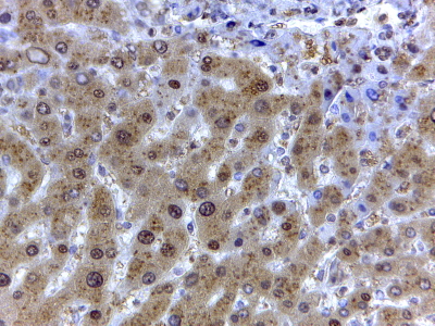
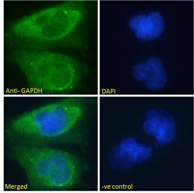
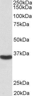
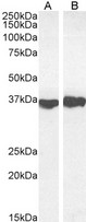
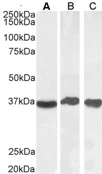
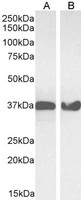
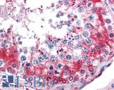
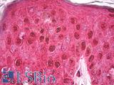
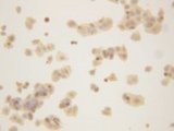
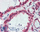
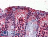
Polyclonal Goat anti‑Human GAPDH Antibody (aa92‑103, IHC, IF, WB) LS‑C20161
Polyclonal Goat anti‑Human GAPDH Antibody (aa92‑103, IHC, IF, WB) LS‑C20161

Note: This antibody replaces LS-C66763
Antibody:
GAPDH Goat anti-Human Polyclonal (aa92-103) Antibody
Application:
IHC-P, IF, WB, Peptide-ELISA
Reactivity:
Human, Monkey, Mouse, Rat, Dog, Hamster, Pig, Rabbit
Format:
Unconjugated, Unmodified
Toll Free North America
 206-374-1102
For Research Use Only
Overview
Antibody:
GAPDH Goat anti-Human Polyclonal (aa92-103) Antibody
Application:
IHC-P, IF, WB, Peptide-ELISA
Reactivity:
Human, Monkey, Mouse, Rat, Dog, Hamster, Pig, Rabbit
Format:
Unconjugated, Unmodified
Specifications
Description
GAPDH antibody LS-C20161 is an unconjugated goat polyclonal antibody to GAPDH (aa92-103) from human. It is reactive with human, mouse, rat and other species. Validated for IF, IHC, Peptide-ELISA and WB. Cited in 3 publications.
Target
Human GAPDH
Synonyms
GAPDH | A1 40 kd subunit | Activator 1 40 kd subunit | G3PD | GAPD | G3pdh | Rfc40 | Rf-c 40 kd subunit
Host
Goat
Reactivity
Human, Monkey, Mouse, Rat, Dog, Hamster, Pig, Rabbit
(tested or 100% immunogen sequence identity)
Clonality
Polyclonal
Conjugations
Unconjugated
Purification
Purified from goat serum by ammonium sulphate precipitation followed by antigen affinity chromatography using the immunizing peptide.
Modifications
Unmodified
Immunogen
Peptide with sequence C-GVNHEKYDNSLK, from the internal region of the protein sequence according to NP_002037.2NP_001243728.1.
Epitope
aa92-103
Specificity
Human GAPDH. GAPDH is constitutively expressed in almost all tissues at high levels. It is therefore a useful marker when a loading/positive control is required in western blotting.
Applications
- IHC - Paraffin (2 µg/ml)
- Immunofluorescence (5 - 10 µg/ml)
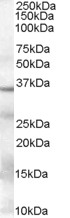
- Western blot (0.03 - 0.1 µg/ml)
- Peptide Enzyme-Linked Immunosorbent Assay (1:16000)

|
Performing IHC? See our complete line of Immunohistochemistry Reagents including antigen retrieval solutions, blocking agents
ABC Detection Kits and polymers, biotinylated secondary antibodies, substrates and more.
|
Presentation
TBS, pH 7.3, 0.02% Sodium Azide, 0.5% BSA
Storage
Aliquot and store at -20°C. Avoid freeze-thaw cycles.
Restrictions
For research use only. Intended for use by laboratory professionals.
About GAPDH
LSBio Ratings
GAPDH Antibody (aa92-103) for IHC, IF/Immunofluorescence, WB/Western LS-C20161 has an LSBio Rating of
Publications (4.2)
Learn more about The LSBio Ratings Algorithm
Publications (3)
Elucidating the phenomenon of HESC-derived RPE: anatomy of cell genesis, expansion and retinal transplantation. Vugler A, Carr AJ, Lawrence J, Chen LL, Burrell K, Wright A, Lundh P, Semo M, Ahmado A, Gias C, da Cruz L, Moore H, Andrews P, Walsh J, Coffey P. Experimental neurology. 2008 214:347-61.
Molecular characterization and functional analysis of phagocytosis by human embryonic stem cell-derived RPE cells using a novel human retinal assay. Carr AJ, Vugler A, Lawrence J, Chen LL, Ahmado A, Chen FK, Semo M, Gias C, da Cruz L, Moore HD, Walsh J, Coffey PJ. Molecular vision. 2009 15:283-95.
The expression of retinal cell markers in human retinal pigment epithelial cells and their augmentation by the synthetic retinoid fenretinide. Carr AJ, Vugler AA, Yu L, Semo M, Coffey P, Moss SE, Greenwood J. Molecular vision. 2011 17:1701-15.
Customer Reviews (0)
Featured Products
Species:
Human, Monkey
Applications:
IHC, IHC - Paraffin, Western blot
Species:
Human, Mouse, Rat, Bovine, Cat, Dog, Goat, Pig, Rabbit, Fish
Applications:
IHC, IHC - Paraffin, Immunofluorescence, Western blot, Immunoprecipitation, ELISA
Species:
Human, Mouse, Rat
Applications:
Immunoprecipitation
Species:
Human, Mouse, Rat, Drosophila
Applications:
IHC, IHC - Paraffin, ICC, Immunofluorescence, Western blot
Species:
Human, Mouse, Rat
Applications:
IHC, IHC - Paraffin, Immunofluorescence, Western blot, ELISA

Species:
Pig, Human, Mouse, Rat
Applications:
IHC, IHC - Paraffin, Immunofluorescence, Western blot
Request SDS/MSDS
To request an SDS/MSDS form for this product, please contact our Technical Support department at:
Technical.Support@LSBio.com
Requested From: United States
Date Requested: 4/24/2024
Date Requested: 4/24/2024


