Login
Registration enables users to use special features of this website, such as past
order histories, retained contact details for faster checkout, review submissions, and special promotions.
order histories, retained contact details for faster checkout, review submissions, and special promotions.
Forgot password?
Registration enables users to use special features of this website, such as past
order histories, retained contact details for faster checkout, review submissions, and special promotions.
order histories, retained contact details for faster checkout, review submissions, and special promotions.
Quick Order
Products
Antibodies
ELISA and Assay Kits
Research Areas
Infectious Disease
Resources
Purchasing
Reference Material
Contact Us
Locations
Orders Processing,
Shipping & Receiving,
Warehouse
2 Shaker Rd Suites
B001/B101
Shirley, MA 01464
Production Lab
Floor 6, Suite 620
20700 44th Avenue W
Lynnwood, WA 98036
Telephone Numbers
Tel: +1 (206) 374-1102
Fax: +1 (206) 577-4565
Contact Us
Additional Contact Details
Login
Registration enables users to use special features of this website, such as past
order histories, retained contact details for faster checkout, review submissions, and special promotions.
order histories, retained contact details for faster checkout, review submissions, and special promotions.
Forgot password?
Registration enables users to use special features of this website, such as past
order histories, retained contact details for faster checkout, review submissions, and special promotions.
order histories, retained contact details for faster checkout, review submissions, and special promotions.
Quick Order
| Catalog Number | Size | Price |
|---|---|---|
| LS-B209-100 | 100 µg | $535 |
1 of 2
2 of 2
IHC‑plus™ Polyclonal Rabbit anti‑Mammal Collagen I Antibody (Biotin, IHC, WB) LS‑B209
IHC‑plus™ Polyclonal Rabbit anti‑Mammal Collagen I Antibody (Biotin, IHC, WB) LS‑B209

Note: This antibody replaces LS-C18863
Antibody:
Collagen I Rabbit anti-Mammal Polyclonal (Biotin) Antibody
Application:
IHC, IHC-P, WB, IP, Flo, ELISA
Reactivity:
Mammal, Human, Mouse, Rat, Bovine
Format:
Biotin, Unmodified
Toll Free North America
 206-374-1102
For Research Use Only
Overview
Antibody:
Collagen I Rabbit anti-Mammal Polyclonal (Biotin) Antibody
Application:
IHC, IHC-P, WB, IP, Flo, ELISA
Reactivity:
Mammal, Human, Mouse, Rat, Bovine
Format:
Biotin, Unmodified
Specifications
Description
Collagen I antibody LS-B209 is a biotin-conjugated rabbit polyclonal antibody to Collagen I from mammal. It is reactive with human, mouse, rat and other species. Validated for ELISA, Flow, IHC, IP and WB.
Host
Rabbit
Reactivity
Mammal, Human, Mouse, Rat, Bovine
(tested or 100% immunogen sequence identity)
Clonality
IgG
Polyclonal
Conjugations
Biotin
Purification
Affinity chromatography
Modifications
Unmodified
Immunogen
Collagen Type I from human and bovine placenta.
Specificity
This product has been prepared by immunoaffinity chromatography using immobilized antigens followed by extensive cross-adsorption against other collagens, human serum proteins and non-collagen extracellular matrix proteins to remove any unwanted specificities. Typically negligible cross reactivity against other types of collagens was detected by ELISA against purified standards. Some class specific anti-collagens may be specific for three-dimensional epitopes which may result in diminished reactivity with denatured collagen or formalin-fixed, paraffin embedded tissues. This antibody reacts with most mammalian Type I collagens and has negligible cross-reactivity with Type II, III, IV, V and VI collagens. Non-specific cross-reaction of anti-collagen antibodies with other human serum proteins or non-collagen extracellular matrix proteins is negligible.
Applications
- IHC
- IHC - Paraffin (10 µg/ml)
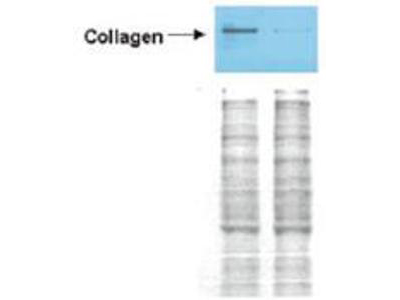
- Western blot (1:3000 - 1:6000)
- Immunoprecipitation (1:100)
- Flow Cytometry
- ELISA (1:3000 - 1:6000)

|
Performing IHC? See our complete line of Immunohistochemistry Reagents including antigen retrieval solutions, blocking agents
ABC Detection Kits and polymers, biotinylated secondary antibodies, substrates and more.
|
Usage
Immunohistochemistry: LS-B209 was validated for use in immunohistochemistry on a panel of 21 formalin-fixed, paraffin-embedded (FFPE) human tissues after heat induced antigen retrieval in pH 6.0 citrate buffer. After incubation with the primary antibody, slides were incubated with biotinylated secondary antibody, followed by alkaline phosphatase-streptavidin and chromogen. The stained slides were evaluated by a pathologist to confirm staining specificity. The optimal working concentration for LS-B209 was determined to be 10 ug/ml. The applications listed have been tested for the unconjugated form of this product. Other forms have not been tested.
Presentation
Lyophilized from 10 mg/mL BSA (Protease and Ig free), 0.02 M Potassium Phosphate, pH 7.2, 0.15 M NaCl, 0.01% Sodium Azide
Reconstitution
Reconstitute in 100 µL of deionized water.
Storage
Store vial at 4°C prior to restoration. For extended storage aliquot contents and freeze at -20°C or below. Avoid freeze-thaw cycles. Stable for several weeks at 4°C as an undiluted liquid. Dilute only prior to immediate use.
Restrictions
For research use only. Intended for use by laboratory professionals.
Validation
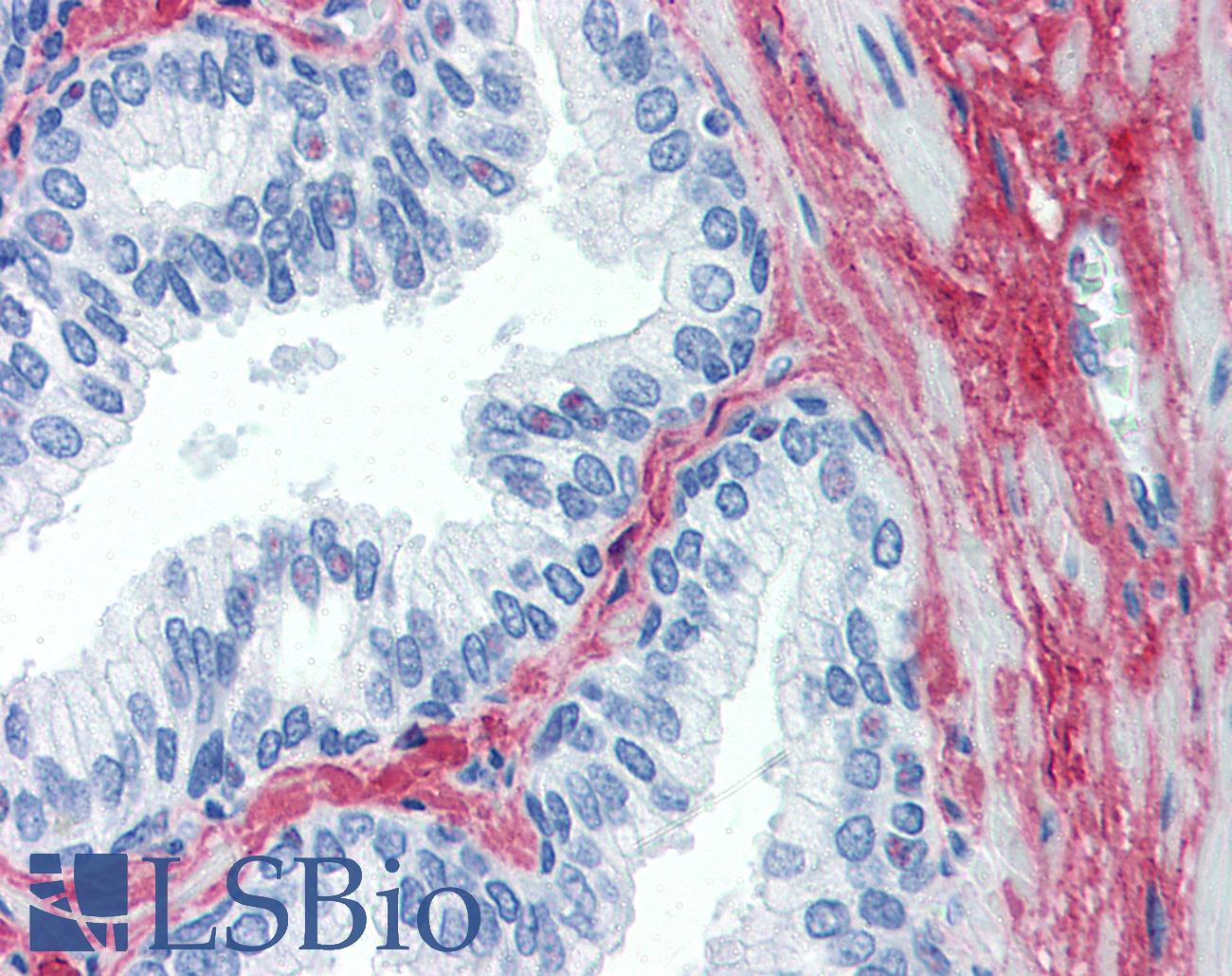
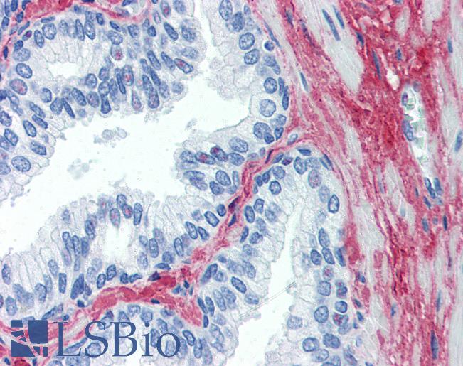
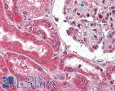
Anti-Collagen I antibody IHC of human prostate. Immunohistochemistry of formalin-fixed, paraffin-embedded tissue after heat-induced antigen retrieval. Antibody concentration 10 ug/ml. This image was taken for the unconjugated form of this product. Other forms have not been tested.
Anti-Collagen I antibody IHC of human prostate. Immunohistochemistry of formalin-fixed, paraffin-embedded tissue after heat-induced antigen retrieval. Antibody concentration 10 ug/ml. This image was taken for the unconjugated form of this product. Other forms have not been tested.
See More About...
LSBio Ratings
IHC-plus™ Collagen I Antibody (Biotin) for IHC, WB/Western, IP, Flow, ELISA LS-B209 has an LSBio Rating of
Laboratory Validation Score (4)
Learn more about The LSBio Ratings Algorithm
Publications (0)
Customer Reviews (0)
Featured Products
Species:
Bovine, Human, Rat, Pig, Rabbit, Deer
Applications:
IHC, IHC - Paraffin, IHC - Frozen, IHC - 4% paraformaldehyde, Immunofluorescence, ELISA
Species:
Human, Sheep
Applications:
IHC, IHC - Frozen, Western blot, Flow Cytometry, ELISA
Species:
Mouse
Applications:
IHC, IHC - Paraffin, IHC - Frozen, Immunofluorescence, Western blot, ELISA, Radioimmunoassay
Species:
Human
Applications:
IHC, IHC - Paraffin, Immunofluorescence, ELISA
Species:
Mouse
Applications:
IHC, IHC - Frozen, Western blot, ELISA, Radioimmunoassay
Request SDS/MSDS
To request an SDS/MSDS form for this product, please contact our Technical Support department at:
Technical.Support@LSBio.com
Requested From: United States
Date Requested: 4/18/2024
Date Requested: 4/18/2024


