Login
Registration enables users to use special features of this website, such as past
order histories, retained contact details for faster checkout, review submissions, and special promotions.
order histories, retained contact details for faster checkout, review submissions, and special promotions.
Forgot password?
Registration enables users to use special features of this website, such as past
order histories, retained contact details for faster checkout, review submissions, and special promotions.
order histories, retained contact details for faster checkout, review submissions, and special promotions.
Quick Order
Products
Antibodies
ELISA and Assay Kits
Research Areas
Infectious Disease
Resources
Purchasing
Reference Material
Contact Us
Locations
Orders Processing,
Shipping & Receiving,
Warehouse
2 Shaker Rd Suites
B001/B101
Shirley, MA 01464
Production Lab
Floor 6, Suite 620
20700 44th Avenue W
Lynnwood, WA 98036
Telephone Numbers
Tel: +1 (206) 374-1102
Fax: +1 (206) 577-4565
Contact Us
Additional Contact Details
Login
Registration enables users to use special features of this website, such as past
order histories, retained contact details for faster checkout, review submissions, and special promotions.
order histories, retained contact details for faster checkout, review submissions, and special promotions.
Forgot password?
Registration enables users to use special features of this website, such as past
order histories, retained contact details for faster checkout, review submissions, and special promotions.
order histories, retained contact details for faster checkout, review submissions, and special promotions.
Quick Order
| Catalog Number | Size | Price |
|---|---|---|
| LS-C154023-100 | 100 µg (1.15 mg/ml) | $567 |
1 of 2
2 of 2
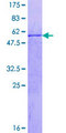
Polyclonal Rabbit anti‑Human CENPQ Antibody (IF, WB) LS‑C154023
Polyclonal Rabbit anti‑Human CENPQ Antibody (IF, WB) LS‑C154023
Antibody:
CENPQ Rabbit anti-Human Polyclonal Antibody
Application:
IF, WB, ELISA
Reactivity:
Human
Format:
Unconjugated, Unmodified
Toll Free North America
 206-374-1102
For Research Use Only
Overview
Antibody:
CENPQ Rabbit anti-Human Polyclonal Antibody
Application:
IF, WB, ELISA
Reactivity:
Human
Format:
Unconjugated, Unmodified
Specifications
Description
CENPQ antibody LS-C154023 is an unconjugated rabbit polyclonal antibody to human CENPQ. Validated for ELISA, IF and WB.
Target
Human CENPQ
Synonyms
CENPQ | C6orf139 | Centromere protein Q | CENP-Q
Host
Rabbit
Reactivity
Human
(tested or 100% immunogen sequence identity)
Clonality
IgG
Polyclonal
Conjugations
Unconjugated
Purification
Protein A purified
Modifications
Unmodified
Immunogen
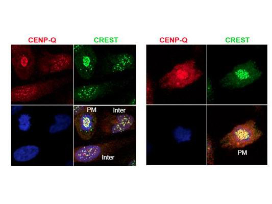
This protein A purified antibody was prepared from whole rabbit serum produced by repeated immunizations with full-length human CENP-Q recombinant protein.
Applications
- Immunofluorescence
- Western blot (1:100 - 1:500)
- ELISA (1:5000)
Usage
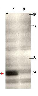
This protein A purified antibody has been tested for use in ELISA, immunofluorescence microscopy and western blotting. Specific conditions for reactivity should be optimized by the end user. Expect a band approximately 26-31 kD in size corresponding to human CENP-Q by western blotting in the appropriate cell lysate or extract.
Presentation
0.02 M Potassium Phosphate, pH 7.2, 0.15 M NaCl, 0.01% Sodium Azide
Storage
Short term: store at 4°C. Long term: aliquot and store at -20°C. Avoid freeze-thaw cycles.
Restrictions
For research use only. Intended for use by laboratory professionals.
About CENPQ
Publications (0)
Customer Reviews (0)
Featured Products
Species:
Human
Applications:
Immunofluorescence, Western blot, ELISA
Request SDS/MSDS
To request an SDS/MSDS form for this product, please contact our Technical Support department at:
Technical.Support@LSBio.com
Requested From: United States
Date Requested: 4/16/2024
Date Requested: 4/16/2024


