Login
Registration enables users to use special features of this website, such as past
order histories, retained contact details for faster checkout, review submissions, and special promotions.
order histories, retained contact details for faster checkout, review submissions, and special promotions.
Forgot password?
Registration enables users to use special features of this website, such as past
order histories, retained contact details for faster checkout, review submissions, and special promotions.
order histories, retained contact details for faster checkout, review submissions, and special promotions.
Quick Order
Products
Antibodies
ELISA and Assay Kits
Research Areas
Infectious Disease
Resources
Purchasing
Reference Material
Contact Us
Locations
Orders Processing,
Shipping & Receiving,
Warehouse
2 Shaker Rd Suites
B001/B101
Shirley, MA 01464
Production Lab
Floor 6, Suite 620
20700 44th Avenue W
Lynnwood, WA 98036
Telephone Numbers
Tel: +1 (206) 374-1102
Fax: +1 (206) 577-4565
Contact Us
Additional Contact Details
Login
Registration enables users to use special features of this website, such as past
order histories, retained contact details for faster checkout, review submissions, and special promotions.
order histories, retained contact details for faster checkout, review submissions, and special promotions.
Forgot password?
Registration enables users to use special features of this website, such as past
order histories, retained contact details for faster checkout, review submissions, and special promotions.
order histories, retained contact details for faster checkout, review submissions, and special promotions.
Quick Order
| Catalog Number | Size | Price |
|---|---|---|
| LS-B10380-50 | 50 µl (1 mg/ml) | $460 |
1 of 4
2 of 4
3 of 4
4 of 4
IHC‑plus™ Monoclonal Mouse anti‑Human VILIP / VSNL1 Antibody (clone 2D11, IHC, IF, WB) LS‑B10380
IHC‑plus™ Monoclonal Mouse anti‑Human VILIP / VSNL1 Antibody (clone 2D11, IHC, IF, WB) LS‑B10380

Note: This antibody replaces LS-C204592
Antibody:
VILIP / VSNL1 Mouse anti-Human Monoclonal (2D11) Antibody
Application:
IHC, IHC-P, IHC-Fr, IF, WB
Reactivity:
Human, Mouse, Rat, Bovine
Format:
Unconjugated, Unmodified
Toll Free North America
 206-374-1102
For Research Use Only
Overview
Antibody:
VILIP / VSNL1 Mouse anti-Human Monoclonal (2D11) Antibody
Application:
IHC, IHC-P, IHC-Fr, IF, WB
Reactivity:
Human, Mouse, Rat, Bovine
Format:
Unconjugated, Unmodified
Specifications
Description
VSNL1 antibody LS-B10380 is an unconjugated mouse monoclonal antibody to VSNL1 (VILIP) from human. It is reactive with human, mouse, rat and other species. Validated for IF, IHC and WB. Tested on 20 paraffin-embedded human tissues.
Target
Human VILIP / VSNL1
Synonyms
VSNL1 | Hippocalcin-like protein 3 | HLP3 | HUVISL1 | HPCAL3 | Visinin-like 1 | VISL1 | VILIP | VILIP-1 | Visinin-like protein 1 | VLP-1
Host
Mouse
Reactivity
Human, Mouse, Rat, Bovine
(tested or 100% immunogen sequence identity)
Clonality
IgG1
Monoclonal
Clone
2D11
Conjugations
Unconjugated
Purification
Affinity purified
Modifications
Unmodified
Immunogen
Recombinant full length human Visinin-like protein 1
Applications
- IHC
- IHC - Paraffin (10 µg/ml)
- IHC - Frozen (1:500 - 1:1000)
- Immunofluorescence (1:500 - 1:1000)
- Western blot (1:1000 - 1:2000)

|
Performing IHC? See our complete line of Immunohistochemistry Reagents including antigen retrieval solutions, blocking agents
ABC Detection Kits and polymers, biotinylated secondary antibodies, substrates and more.
|
Usage
The antibody solution can be used at dilutions of 1:500-1:1000 in immunofluorescence experiments. In western blotting using chemiluminescence it can be used at dilutions of 1:1000-1:2000.
Presentation
PBS, 10 mM Sodium Azide
Storage
Store at 4°C or -20°C. Avoid freeze-thaw cycles.
Restrictions
For research use only. Intended for use by laboratory professionals.
About VILIP / VSNL1
Validation
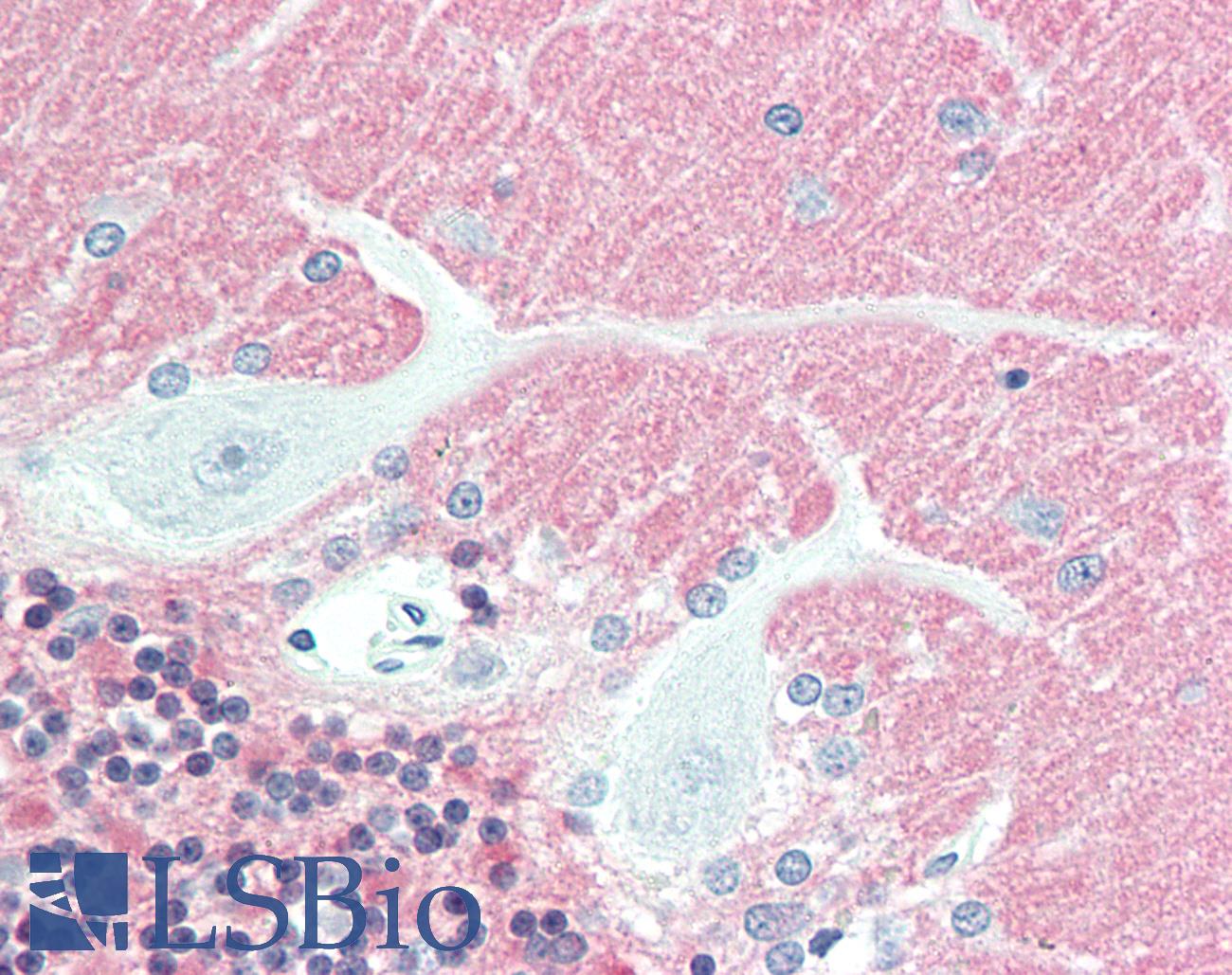
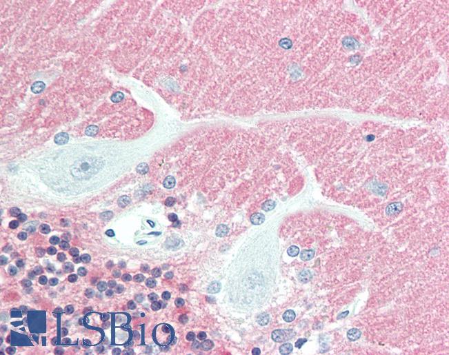
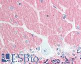
Anti-VILIP / VSNL1 antibody IHC staining of human brain, cerebellum. Immunohistochemistry of formalin-fixed, paraffin-embedded tissue after heat-induced antigen retrieval. Antibody concentration 10 ug/ml.
Anti-VILIP / VSNL1 antibody IHC staining of human brain, cerebellum. Immunohistochemistry of formalin-fixed, paraffin-embedded tissue after heat-induced antigen retrieval. Antibody concentration 10 ug/ml.

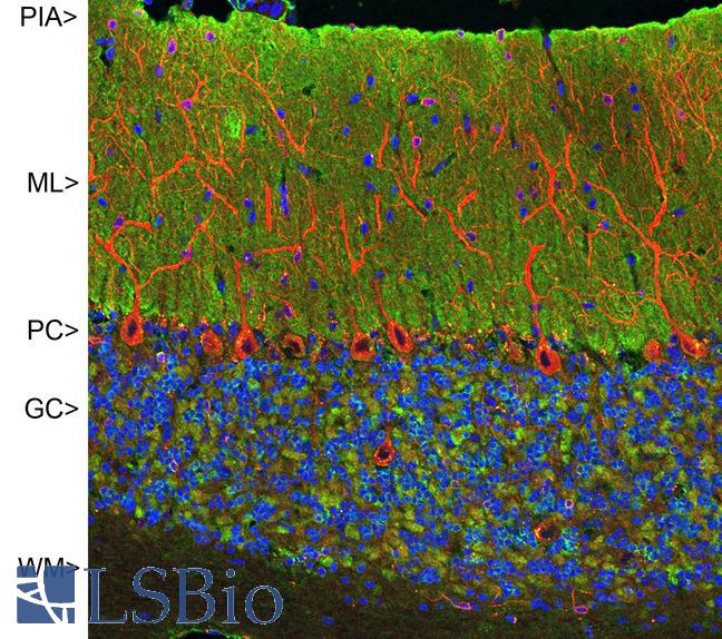
Confocal image of adult rat cerebellar cortex stained with VILIP / VSNL1 antibody (green), chicken polyclonal antibody to MAP2 (red) and DNA (blue). The VILIP / VSNL1 antibody antibody reveals synapses in the molecular layer (ML) strongly. Synaptic regions are also seen in the granule cell layer (GC). The perikarya of Purkinje cells (PC) are revealed with MAP2 antibody (4). Little staining is seen in the white matter (WM).
Confocal image of adult rat cerebellar cortex stained with VILIP / VSNL1 antibody (green), chicken polyclonal antibody to MAP2 (red) and DNA (blue). The VILIP / VSNL1 antibody antibody reveals synapses in the molecular layer (ML) strongly. Synaptic regions are also seen in the granule cell layer (GC). The perikarya of Purkinje cells (PC) are revealed with MAP2 antibody (4). Little staining is seen in the white matter (WM).
See More About...
LSBio Ratings
IHC-plus™ VILIP / VSNL1 Antibody (clone 2D11) for IHC, IF/Immunofluorescence, WB/Western LS-B10380 has an LSBio Rating of
Laboratory Validation Score (4)
Learn more about The LSBio Ratings Algorithm
Publications (0)
Customer Reviews (0)
Featured Products
Species:
Human, Mouse, Rat, Bovine
Applications:
IHC, IHC - Paraffin, Immunofluorescence, Western blot
Species:
Human, Mouse
Applications:
IHC, IHC - Paraffin, Western blot
Request SDS/MSDS
To request an SDS/MSDS form for this product, please contact our Technical Support department at:
Technical.Support@LSBio.com
Requested From: United States
Date Requested: 4/19/2024
Date Requested: 4/19/2024


