Login
Registration enables users to use special features of this website, such as past
order histories, retained contact details for faster checkout, review submissions, and special promotions.
order histories, retained contact details for faster checkout, review submissions, and special promotions.
Forgot password?
Registration enables users to use special features of this website, such as past
order histories, retained contact details for faster checkout, review submissions, and special promotions.
order histories, retained contact details for faster checkout, review submissions, and special promotions.
Quick Order
Products
Antibodies
ELISA and Assay Kits
Research Areas
Infectious Disease
Resources
Purchasing
Reference Material
Contact Us
Locations
Orders Processing,
Shipping & Receiving,
Warehouse
2 Shaker Rd Suites
B001/B101
Shirley, MA 01464
Production Lab
Floor 6, Suite 620
20700 44th Avenue W
Lynnwood, WA 98036
Telephone Numbers
Tel: +1 (206) 374-1102
Fax: +1 (206) 577-4565
Contact Us
Additional Contact Details
Login
Registration enables users to use special features of this website, such as past
order histories, retained contact details for faster checkout, review submissions, and special promotions.
order histories, retained contact details for faster checkout, review submissions, and special promotions.
Forgot password?
Registration enables users to use special features of this website, such as past
order histories, retained contact details for faster checkout, review submissions, and special promotions.
order histories, retained contact details for faster checkout, review submissions, and special promotions.
Quick Order
| Catalog Number | Size | Price |
|---|---|---|
| LS-B10591-50 | 50 µg (1 mg/ml) | $485 |
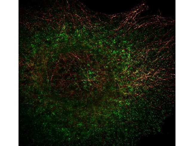
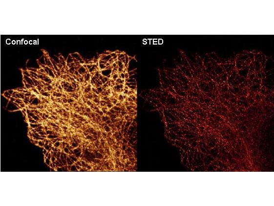
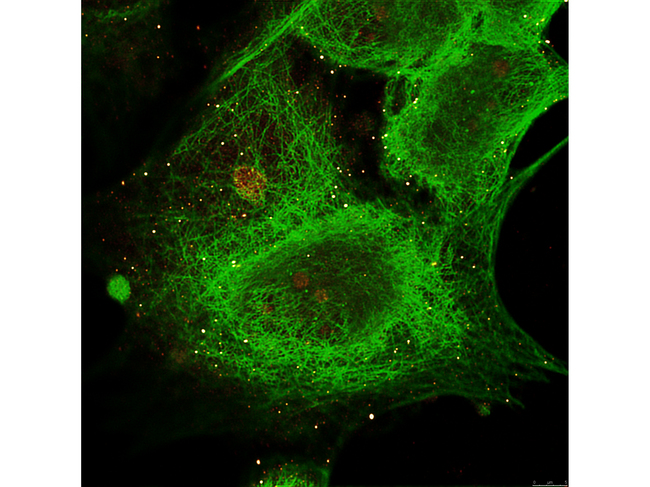
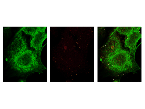
![TUBA1B / Tubulin Alpha 1B Antibody - Immunofluorescence Microscopy - alpha-Tubulin Monoclonal Antibody -. a-tubulin monoclonal antibody detects tubulin (colored RED) in STED immunofluorescence microscopy. Methanol fixed A431 cells were blocked with normal goat serum. The cells were then probed with 0.4 ug/mL final concentration of anti-a-tubulin and detected with 0.2 ug/mL ATTO 425 conjugated anti-MOUSE IgG [GOAT] ( secondary antibody. Also shown in this 2-color STED image is Anti-HDAC-1 [RABBIT] (LS-B34) detected with DyLight488 conjugated Anti-RABBIT IgG [GOAT] secondary antibody (colored GREEN). Image courtesy of Myriam Gastard, Leica Microsystems, USA.](https://lsbio-7d62.kxcdn.com/image2/ihc-plus-tuba1b-tubulin-alpha-1b-antibody-c-terminus-clone-17h11.f10-ls-b10591/81768_38342.jpg)
1 of 8
2 of 8
3 of 8
4 of 8
5 of 8
6 of 8
7 of 8
8 of 8
IHC‑plus™ Monoclonal Mouse anti‑Human TUBA1B / Tubulin Alpha 1B Antibody (clone 17H11.F10, C‑Terminus, IHC, IF, WB) LS‑B10591
IHC‑plus™ Monoclonal Mouse anti‑Human TUBA1B / Tubulin Alpha 1B Antibody (clone 17H11.F10, C‑Terminus, IHC, IF, WB) LS‑B10591

Note: This antibody replaces LS-C154003
Antibody:
TUBA1B / Tubulin Alpha 1B Mouse anti-Human Monoclonal (C-Terminus) (17H11.F10) Antibody
Application:
IHC, IHC-P, IF, WB, ELISA
Reactivity:
Human, Mouse, Rat, Bovine, Chicken
Format:
Unconjugated, Unmodified
Toll Free North America
 206-374-1102
For Research Use Only
Overview
Antibody:
TUBA1B / Tubulin Alpha 1B Mouse anti-Human Monoclonal (C-Terminus) (17H11.F10) Antibody
Application:
IHC, IHC-P, IF, WB, ELISA
Reactivity:
Human, Mouse, Rat, Bovine, Chicken
Format:
Unconjugated, Unmodified
Specifications
Description
Tubulin Alpha 1B antibody LS-B10591 is an unconjugated mouse monoclonal antibody to Tubulin Alpha 1B (TUBA1B) (C-Terminus) from human. It is reactive with human, mouse, rat and other species. Validated for ELISA, IF, IHC and WB. Tested on 20 paraffin-embedded human tissues.
Target
Human TUBA1B / Tubulin Alpha 1B
Synonyms
TUBA1B | Alpha-tubulin ubiquitous | Alpha tubulin | K-ALPHA-1 | Tubulin, alpha 1b | Tubulin alpha-ubiquitous chain | Tubulin, alpha, ubiquitous | Tubulin K-alpha-1 | Tubulin alpha chain | Tubulin alpha-1B chain
Host
Mouse
Reactivity
Human, Mouse, Rat, Bovine, Chicken
(tested or 100% immunogen sequence identity)
Predicted
Fish, Algae, Avian, Bacteria, Mammal, Protozoa (at least 90% immunogen sequence identity)
Clonality
IgG1,k
Monoclonal
Clone
17H11.F10
Conjugations
Unconjugated
Purification
Protein A purified
Modifications
Unmodified
Immunogen
Anti-Tubulin Loading Control Antibody was produced by repeated immunizations with a synthetic peptide corresponding to residues near the C terminal end of human alpha tubulin protein.
Epitope
C-Terminus
Specificity
A BLAST analysis was used to suggest antibody reactivity with alpha tubulin from a wide range of organisms, including avian, mammalian aquatic, parasitic and alga sources based on 100% homology for the immunogen sequence. Cross reactivity will occur with all isoforms of alpha tubulin. Such broad reactivity makes this antibody useful as an excellent loading control.
Applications
- IHC
- IHC - Paraffin (5 µg/ml)
- Immunofluorescence (0.1 µg/ml)
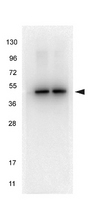
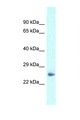
- Western blot (1:1000)
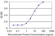
- ELISA (1:300000)

|
Performing IHC? See our complete line of Immunohistochemistry Reagents including antigen retrieval solutions, blocking agents
ABC Detection Kits and polymers, biotinylated secondary antibodies, substrates and more.
|
Usage
Anti-Tubulin Antibody has been tested for use in ELISA, immunohistochemistry, immunofluorescence microscopy and western blot. Specific conditions for reactivity should be optimized by the end user. Expect a band at ~50 kD in size corresponding to alpha tubulin by western blotting in most cell lysates or extracts.
Presentation
0.02 M Potassium Phosphate, pH 7.2, 0.15 M NaCl, 0.01% Sodium Azide
Storage
Short term: store at 4°C. Long term: aliquot and store at -20°C. Avoid freeze-thaw cycles.
Restrictions
For research use only. Intended for use by laboratory professionals.
About TUBA1B / Tubulin Alpha 1B
Validation

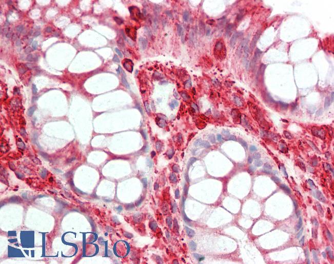
Anti-TUBA1B antibody IHC staining of human colon. Immunohistochemistry of formalin-fixed, paraffin-embedded tissue after heat-induced antigen retrieval. Antibody concentration 5 ug/ml.
Anti-TUBA1B antibody IHC staining of human colon. Immunohistochemistry of formalin-fixed, paraffin-embedded tissue after heat-induced antigen retrieval. Antibody concentration 5 ug/ml.
See More About...
LSBio Ratings
IHC-plus™ TUBA1B / Tubulin Alpha 1B Antibody (C-Terminus, clone 17H11.F10) for IHC, IF/Immunofluorescence, WB/Western, ELISA LS-B10591 has an LSBio Rating of
Laboratory Validation Score (4)
Learn more about The LSBio Ratings Algorithm
Publications (0)
Customer Reviews (0)
Featured Products
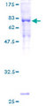
Source:
Wheat
Tag:
GST
Reactivity:
Human
Range:
0.78-50 ng/ml
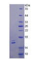
Source:
E. coli
Tag:
6His, C-terminus
Source:
E. coli
Tag:
His
Request SDS/MSDS
To request an SDS/MSDS form for this product, please contact our Technical Support department at:
Technical.Support@LSBio.com
Requested From: United States
Date Requested: 4/19/2024
Date Requested: 4/19/2024


