Login
Registration enables users to use special features of this website, such as past
order histories, retained contact details for faster checkout, review submissions, and special promotions.
order histories, retained contact details for faster checkout, review submissions, and special promotions.
Forgot password?
Registration enables users to use special features of this website, such as past
order histories, retained contact details for faster checkout, review submissions, and special promotions.
order histories, retained contact details for faster checkout, review submissions, and special promotions.
Quick Order
Products
Antibodies
ELISA and Assay Kits
Research Areas
Infectious Disease
Resources
Purchasing
Reference Material
Contact Us
Locations
Orders Processing,
Shipping & Receiving,
Warehouse
2 Shaker Rd Suites
B001/B101
Shirley, MA 01464
Production Lab
Floor 6, Suite 620
20700 44th Avenue W
Lynnwood, WA 98036
Telephone Numbers
Tel: +1 (206) 374-1102
Fax: +1 (206) 577-4565
Contact Us
Additional Contact Details
Login
Registration enables users to use special features of this website, such as past
order histories, retained contact details for faster checkout, review submissions, and special promotions.
order histories, retained contact details for faster checkout, review submissions, and special promotions.
Forgot password?
Registration enables users to use special features of this website, such as past
order histories, retained contact details for faster checkout, review submissions, and special promotions.
order histories, retained contact details for faster checkout, review submissions, and special promotions.
Quick Order
| Catalog Number | Size | Price |
|---|---|---|
| LS-B431-50 | 50 µg (1 mg/ml) | $515 |
1 of 5
2 of 5
3 of 5
4 of 5
5 of 5
IHC‑plus™ Monoclonal Mouse anti‑Human TP53 / p53 Antibody (clone BP53‑12, IHC, IF, WB) LS‑B431
IHC‑plus™ Monoclonal Mouse anti‑Human TP53 / p53 Antibody (clone BP53‑12, IHC, IF, WB) LS‑B431

Note: This antibody replaces LS-C19012
Antibody:
TP53 / p53 Mouse anti-Human Monoclonal (BP53-12) Antibody
Application:
IHC, IHC-P, IF, WB, IP, ELISA, ChrIP
Reactivity:
Human
Format:
Unconjugated, Unmodified
Toll Free North America
 206-374-1102
For Research Use Only
Overview
Antibody:
TP53 / p53 Mouse anti-Human Monoclonal (BP53-12) Antibody
Application:
IHC, IHC-P, IF, WB, IP, ELISA, ChrIP
Reactivity:
Human
Format:
Unconjugated, Unmodified
Specifications
Description
P53 antibody LS-B431 is an unconjugated mouse monoclonal antibody to human p53 (TP53). Validated for ChrIP, ELISA, IF, IHC, IP and WB. Tested on 20 paraffin-embedded human tissues. Cited in 3 publications.
Target
Human TP53 / p53
Synonyms
TP53 | BCC7 | Cellular tumor antigen p53 | p53 | TRP53 | p53 tumor suppressor | Phosphoprotein p53 | Antigen NY-CO-13 | Tumor protein p53 | Tumor suppressor p53 | LFS1
Host
Mouse
Reactivity
Human
(tested or 100% immunogen sequence identity)
Clonality
IgG2a,k
Monoclonal
Clone
BP53-12
Conjugations
Unconjugated
Purification
Protein A purified
Modifications
Unmodified
Immunogen
This protein A purified monoclonal antibody was produced by repeated immunizations with recombinant human p53 protein.
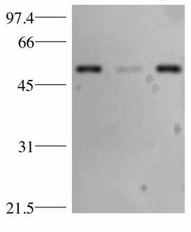
Specificity
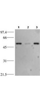
Reacts specifically with p53 in human tissues and cell lines. The antibody recognizes a 53 kDa band corresponding to p53. Cross reactivity with p53 from other sources has not been determined.
Applications
- IHC
- IHC - Paraffin (2.5 µg/ml)
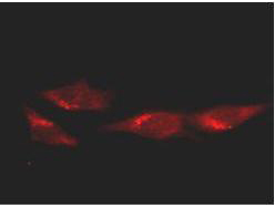
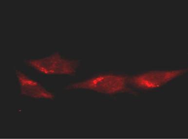
- Immunofluorescence (1:100 - 1:500)
- Western blot (1:500 - 1:2000)
- Immunoprecipitation (1:100)
- ELISA (1:2000 - 1:10000)
- Chromatin Immunoprecipitation (1 µg/µl)

|
Performing IHC? See our complete line of Immunohistochemistry Reagents including antigen retrieval solutions, blocking agents
ABC Detection Kits and polymers, biotinylated secondary antibodies, substrates and more.
|
Usage
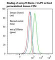
Immunohistochemistry: LS-B431 was validated for use in immunohistochemistry on a panel of 21 formalin-fixed, paraffin-embedded (FFPE) human tissues after heat induced antigen retrieval in pH 6.0 citrate buffer. After incubation with the primary antibody, slides were incubated with biotinylated secondary antibody, followed by alkaline phosphatase-streptavidin and chromogen. The stained slides were evaluated by a pathologist to confirm staining specificity. The optimal working concentration for LS-B431 was determined to be 2.5 ug/ml.
Presentation
0.02 M Potassium Phosphate, pH 7.2, 0.5 M NaCl, 0.01% Sodium Azide
Storage
Store at 4°C or -20°C. Avoid freeze-thaw cycles.
Restrictions
For research use only. Intended for use by laboratory professionals.
About TP53 / p53
Validation

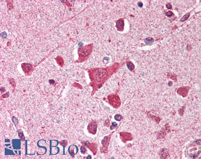
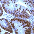
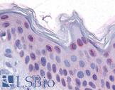
Anti-TP53 / p53 antibody IHC of human brain, cortex. Immunohistochemistry of formalin-fixed, paraffin-embedded tissue after heat-induced antigen retrieval. Antibody concentration 5 ug/ml.
Anti-TP53 / p53 antibody IHC of human brain, cortex. Immunohistochemistry of formalin-fixed, paraffin-embedded tissue after heat-induced antigen retrieval. Antibody concentration 5 ug/ml.
See More About...
LSBio Ratings
IHC-plus™ TP53 / p53 Antibody (clone BP53-12) for IHC, IF/Immunofluorescence, WB/Western, IP, ELISA LS-B431 has an LSBio Rating of
Publications (4.2)
Laboratory Validation Score (4)
Learn more about The LSBio Ratings Algorithm
Publications (3)
p53 mutations in human cancers. Hollstein M, Sidransky D, Vogelstein B, Harris CC. Science (New York, N.Y.). 1991 253:49-53.
Monoclonal antibodies against simian virus 40 T antigens: evidence for distinct sublcasses of large T antigen and for similarities among nonviral T antigens. Gurney EG, Harrison RO, Fenno J. Journal of virology. 1980 34:752-63.
Customer Reviews (0)
Featured Products
Species:
Mouse, Human, Rat, Bovine, Hamster, Chicken
Applications:
IHC, Western blot, Flow Cytometry
Species:
Human
Applications:
IHC, IHC - Paraffin, Western blot
Species:
Human
Applications:
IHC, IHC - Paraffin, IHC - Frozen, Western blot, Immunoprecipitation, ELISA
Species:
Human
Applications:
IHC - Paraffin, IHC - Frozen, Western blot, Immunoprecipitation
Reactivity:
Human, Mouse, Rat
Range:
Positive/Negative
Request SDS/MSDS
To request an SDS/MSDS form for this product, please contact our Technical Support department at:
Technical.Support@LSBio.com
Requested From: United States
Date Requested: 4/16/2024
Date Requested: 4/16/2024


