order histories, retained contact details for faster checkout, review submissions, and special promotions.
Forgot password?
order histories, retained contact details for faster checkout, review submissions, and special promotions.
Locations
Orders Processing,
Shipping & Receiving,
Warehouse
2 Shaker Rd Suites
B001/B101
Shirley, MA 01464
Production Lab
Floor 6, Suite 620
20700 44th Avenue W
Lynnwood, WA 98036
Telephone Numbers
Tel: +1 (206) 374-1102
Fax: +1 (206) 577-4565
Contact Us
Additional Contact Details
order histories, retained contact details for faster checkout, review submissions, and special promotions.
Forgot password?
order histories, retained contact details for faster checkout, review submissions, and special promotions.
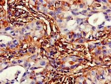
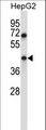
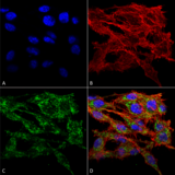
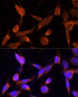
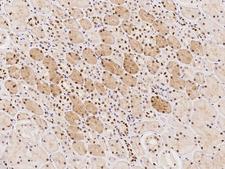
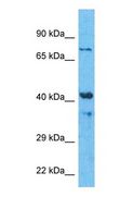
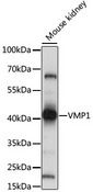
TMEM49
vacuole membrane protein 1
Stress-induced protein that, when overexpressed, promotes formation of intracellular vacuoles followed by cell death. May be involved in the cytoplasmic vacuolization of acinar cells during the early stage of acute pancreatitis. Plays a role in the initial stages of the autophagic process through its interaction with BECN1 By similarity. Involved in cell-cell adhesion. Plays an essential role in formation of cell junctions.
| Gene Name: | vacuole membrane protein 1 |
| Synonyms: | VMP1, TMEM49, TANGO5, Transmembrane protein 49, EPG3, TDC1, Vacuole membrane protein 1 |
| Target Sequences: | NM_030938 NP_112200.2 Q96GC9 |


If you do not find the reagent or information you require, please contact Customer.Support@LSBio.com to inquire about additional products in development.


