Login
Registration enables users to use special features of this website, such as past
order histories, retained contact details for faster checkout, review submissions, and special promotions.
order histories, retained contact details for faster checkout, review submissions, and special promotions.
Forgot password?
Registration enables users to use special features of this website, such as past
order histories, retained contact details for faster checkout, review submissions, and special promotions.
order histories, retained contact details for faster checkout, review submissions, and special promotions.
Quick Order
Products
Antibodies
ELISA and Assay Kits
Research Areas
Infectious Disease
Resources
Purchasing
Reference Material
Contact Us
Locations
Orders Processing,
Shipping & Receiving,
Warehouse
2 Shaker Rd Suites
B001/B101
Shirley, MA 01464
Production Lab
Floor 6, Suite 620
20700 44th Avenue W
Lynnwood, WA 98036
Telephone Numbers
Tel: +1 (206) 374-1102
Fax: +1 (206) 577-4565
Contact Us
Additional Contact Details
Login
Registration enables users to use special features of this website, such as past
order histories, retained contact details for faster checkout, review submissions, and special promotions.
order histories, retained contact details for faster checkout, review submissions, and special promotions.
Forgot password?
Registration enables users to use special features of this website, such as past
order histories, retained contact details for faster checkout, review submissions, and special promotions.
order histories, retained contact details for faster checkout, review submissions, and special promotions.
Quick Order
| Catalog Number | Size | Price |
|---|---|---|
| LS-B3451-200 | 200 µl (1 mg/ml) | $460 |

1 of 6
2 of 6
3 of 6
4 of 6
5 of 6
6 of 6
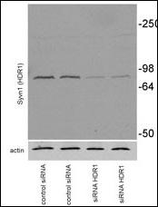
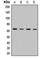
IHC‑plus™ Polyclonal Rabbit anti‑Human SYVN1 / HRD1 Antibody (aa586‑617, IHC, IF, WB) LS‑B3451
IHC‑plus™ Polyclonal Rabbit anti‑Human SYVN1 / HRD1 Antibody (aa586‑617, IHC, IF, WB) LS‑B3451

Note: This antibody replaces LS-C99633
Antibody:
SYVN1 / HRD1 Rabbit anti-Human Polyclonal (aa586-617) Antibody
Application:
IHC, IHC-P, IF, WB
Reactivity:
Human, Mouse
Format:
Unconjugated, Unmodified
Toll Free North America
 206-374-1102
For Research Use Only
Overview
Antibody:
SYVN1 / HRD1 Rabbit anti-Human Polyclonal (aa586-617) Antibody
Application:
IHC, IHC-P, IF, WB
Reactivity:
Human, Mouse
Format:
Unconjugated, Unmodified
Specifications
Description
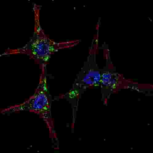
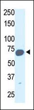
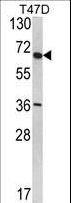
HRD1 antibody LS-B3451 is an unconjugated rabbit polyclonal antibody to HRD1 (SYVN1) (aa586-617) from human. It is reactive with human and mouse. Validated for IF, IHC and WB. Tested on 20 paraffin-embedded human tissues. Cited in 2 publications.
Target
Human SYVN1 / HRD1
Synonyms
SYVN1 | KIAA1810 | Synoviolin | Synovial apoptosis inhibitor 1 | DER3 | HRD1
Host
Rabbit
Reactivity
Human, Mouse
(tested or 100% immunogen sequence identity)
Clonality
Polyclonal
Conjugations
Unconjugated
Purification
Ammonium sulfate precipitation
Modifications
Unmodified
Epitope
aa586-617
Specificity
This SYVN1 (HRD1) antibody is generated from rabbits immunized with a KLH conjugated synthetic peptide between 586-617 amino acids from the C-terminal region of human SYVN1 (HRD1).
Applications
- IHC
- IHC - Paraffin (10 µg/ml)
- Immunofluorescence (1:200)
- Western blot (1:1000)

|
Performing IHC? See our complete line of Immunohistochemistry Reagents including antigen retrieval solutions, blocking agents
ABC Detection Kits and polymers, biotinylated secondary antibodies, substrates and more.
|
Usage
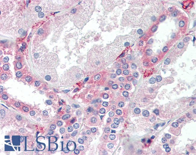
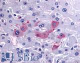
Immunohistochemistry: LS-B3451 was validated for use in immunohistochemistry on a panel of 21 formalin-fixed, paraffin-embedded (FFPE) human tissues after heat induced antigen retrieval in pH 6.0 citrate buffer. After incubation with the primary antibody, slides were incubated with biotinylated secondary antibody, followed by alkaline phosphatase-streptavidin and chromogen. The stained slides were evaluated by a pathologist to confirm staining specificity. The optimal working concentration for LS-B3451 was determined to be 10 ug/ml.
Presentation
PBS, 0.09% Sodium Azide
Storage
Maintain refrigerated at 2°C to 8°C for up to 6 months. For long term storage store at -20°C.
Restrictions
For research use only. Intended for use by laboratory professionals.
About SYVN1 / HRD1
Validation
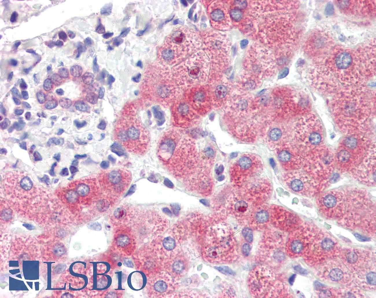
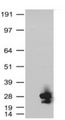
Anti-SYVN1 / HRD1 antibody IHC of human liver. Immunohistochemistry of formalin-fixed, paraffin-embedded tissue after heat-induced antigen retrieval. Antibody concentration 10 ug/ml.
Anti-SYVN1 / HRD1 antibody IHC of human liver. Immunohistochemistry of formalin-fixed, paraffin-embedded tissue after heat-induced antigen retrieval. Antibody concentration 10 ug/ml.
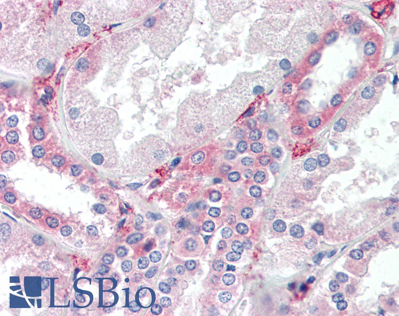
Anti-SYVN1 / HRD1 antibody IHC of human kidney. Immunohistochemistry of formalin-fixed, paraffin-embedded tissue after heat-induced antigen retrieval. Antibody concentration 10 ug/ml.
Anti-SYVN1 / HRD1 antibody IHC of human kidney. Immunohistochemistry of formalin-fixed, paraffin-embedded tissue after heat-induced antigen retrieval. Antibody concentration 10 ug/ml.
See More About...
LSBio Ratings
IHC-plus™ SYVN1 / HRD1 Antibody (aa586-617) for IHC, IF/Immunofluorescence, WB/Western LS-B3451 has an LSBio Rating of
Publications (4.1)
Laboratory Validation Score (4)
Learn more about The LSBio Ratings Algorithm
Publications (2)
Loss of HRD1-mediated protein degradation causes amyloid precursor protein accumulation and amyloid-beta generation. Kaneko M, Koike H, Saito R, Kitamura Y, Okuma Y, Nomura Y. The Journal of neuroscience : the official journal of the Society for Neuroscience. 2010 30:3924-32.
Ubiquitin ligase substrate identification through quantitative proteomics at both the protein and peptide levels. Lee KA, Hammerle LP, Andrews PS, Stokes MP, Mustelin T, Silva JC, Black RA, Doedens JR. The Journal of biological chemistry. 2011 286:41530-8.
Customer Reviews (0)
Featured Products
Species:
Human, Monkey, Mouse, Rat, Bat, Bovine, Dog, Hamster, Horse, Rabbit, Xenopus, Zebrafish
Applications:
IHC, IHC - Paraffin
Species:
Human, Mouse, Rat
Applications:
Western blot, Immunoprecipitation
Reactivity:
Human, Mouse, Rat
Range:
Positive/Negative
Species:
Rabbit, Human, Monkey, Mouse, Rat, Dog, Pig, Xenopus
Applications:
IHC, Western blot, Immunoprecipitation
Request SDS/MSDS
To request an SDS/MSDS form for this product, please contact our Technical Support department at:
Technical.Support@LSBio.com
Requested From: United States
Date Requested: 4/24/2024
Date Requested: 4/24/2024


