Login
Registration enables users to use special features of this website, such as past
order histories, retained contact details for faster checkout, review submissions, and special promotions.
order histories, retained contact details for faster checkout, review submissions, and special promotions.
Forgot password?
Registration enables users to use special features of this website, such as past
order histories, retained contact details for faster checkout, review submissions, and special promotions.
order histories, retained contact details for faster checkout, review submissions, and special promotions.
Quick Order
Products
Antibodies
ELISA and Assay Kits
Research Areas
Infectious Disease
Resources
Purchasing
Reference Material
Contact Us
Locations
Orders Processing,
Shipping & Receiving,
Warehouse
2 Shaker Rd Suites
B001/B101
Shirley, MA 01464
Production Lab
Floor 6, Suite 620
20700 44th Avenue W
Lynnwood, WA 98036
Telephone Numbers
Tel: +1 (206) 374-1102
Fax: +1 (206) 577-4565
Contact Us
Additional Contact Details
Login
Registration enables users to use special features of this website, such as past
order histories, retained contact details for faster checkout, review submissions, and special promotions.
order histories, retained contact details for faster checkout, review submissions, and special promotions.
Forgot password?
Registration enables users to use special features of this website, such as past
order histories, retained contact details for faster checkout, review submissions, and special promotions.
order histories, retained contact details for faster checkout, review submissions, and special promotions.
Quick Order
| Catalog Number | Size | Price |
|---|---|---|
| LS-B10609-50 | 50 µg | $515 |
1 of 4
2 of 4
3 of 4
4 of 4
IHC‑plus™ Polyclonal Rabbit anti‑Mouse SIPA1 Antibody (IHC, WB) LS‑B10609
IHC‑plus™ Polyclonal Rabbit anti‑Mouse SIPA1 Antibody (IHC, WB) LS‑B10609

Note: This antibody replaces LS-C60117
Antibody:
SIPA1 Rabbit anti-Mouse Polyclonal Antibody
Application:
IHC, IHC-P, WB, ELISA
Reactivity:
Mouse, Human, Rat
Format:
Unconjugated, Unmodified
Toll Free North America
 206-374-1102
For Research Use Only
Overview
Antibody:
SIPA1 Rabbit anti-Mouse Polyclonal Antibody
Application:
IHC, IHC-P, WB, ELISA
Reactivity:
Mouse, Human, Rat
Format:
Unconjugated, Unmodified
Specifications
Description
SIPA1 antibody LS-B10609 is an unconjugated rabbit polyclonal antibody to SIPA1 from mouse. It is reactive with human, mouse and rat. Validated for ELISA, IHC and WB.
Host
Rabbit
Reactivity
Mouse, Human, Rat
(tested or 100% immunogen sequence identity)
Predicted
Human, Rat (at least 90% immunogen sequence identity)
Clonality
IgG
Polyclonal
Conjugations
Unconjugated
Purification
Affinity purified
Modifications
Unmodified
Immunogen
This affinity purified antibody was prepared from whole rabbit serum produced by repeated immunizations with a synthetic peptide corresponding to a region near the amino terminus of mouse Sipa1.
Specificity
This antibody is specific for mouse Sipa1 protein. A BLAST analysis was used to suggest cross-reactivity with Sipa1 from mouse, human and rat based on a 100% homology with the immunizing sequence. Cross-reactivity with Sipa1 from other sources has not been determined
Applications
- IHC
- IHC - Paraffin (5 µg/ml)
- Western blot (1:1000 - 1:5000)
- ELISA (1:20000)

|
Performing IHC? See our complete line of Immunohistochemistry Reagents including antigen retrieval solutions, blocking agents
ABC Detection Kits and polymers, biotinylated secondary antibodies, substrates and more.
|
Usage
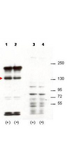
This affinity purified antibody has been tested for use in ELISA, immunohistochemistry and western blotting. Specific conditions for reactivity should be optimized by the end user. Expect a band approximately 130 kD in size corresponding to Sipa1 by western blotting in the appropriate cell lysate or extract. This antibody is capable of detecting both over-expressed and endogenous Sipa1.
Presentation
0.02 M Potassium Phosphate, pH 7.2, 0.15 M NaCl, 0.01% Sodium Azide
Storage
Short term: -20°C; Long term: -20°C.
Restrictions
For research use only. Intended for use by laboratory professionals.
About SIPA1
Validation
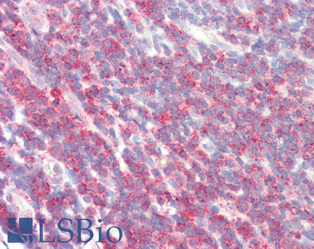
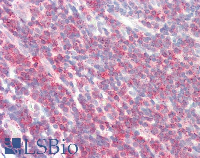
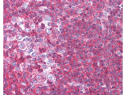
Anti-SIPA1 antibody IHC staining of human tonsil. Immunohistochemistry of formalin-fixed, paraffin-embedded tissue after heat-induced antigen retrieval.
Anti-SIPA1 antibody IHC staining of human tonsil. Immunohistochemistry of formalin-fixed, paraffin-embedded tissue after heat-induced antigen retrieval.

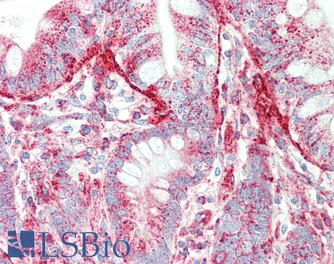
Anti-SIPA1 antibody IHC staining of human small intestine. Immunohistochemistry of formalin-fixed, paraffin-embedded tissue after heat-induced antigen retrieval. Antibody concentration 5 ug/ml.
Anti-SIPA1 antibody IHC staining of human small intestine. Immunohistochemistry of formalin-fixed, paraffin-embedded tissue after heat-induced antigen retrieval. Antibody concentration 5 ug/ml.
See More About...
LSBio Ratings
IHC-plus™ SIPA1 Antibody for IHC, WB/Western, ELISA LS-B10609 has an LSBio Rating of
Laboratory Validation Score (4)
Learn more about The LSBio Ratings Algorithm
Publications (0)
Customer Reviews (0)
Featured Products
Reactivity:
Human
Range:
0.78-50 ng/ml

Reactivity:
Pig
Range:
0.156-10 ng/ml
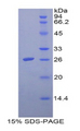
Source:
E. coli
Tag:
His

Request SDS/MSDS
To request an SDS/MSDS form for this product, please contact our Technical Support department at:
Technical.Support@LSBio.com
Requested From: United States
Date Requested: 4/18/2024
Date Requested: 4/18/2024


