order histories, retained contact details for faster checkout, review submissions, and special promotions.
Forgot password?
order histories, retained contact details for faster checkout, review submissions, and special promotions.
Locations
Orders Processing,
Shipping & Receiving,
Warehouse
2 Shaker Rd Suites
B001/B101
Shirley, MA 01464
Production Lab
Floor 6, Suite 620
20700 44th Avenue W
Lynnwood, WA 98036
Telephone Numbers
Tel: +1 (206) 374-1102
Fax: +1 (206) 577-4565
Contact Us
Additional Contact Details
order histories, retained contact details for faster checkout, review submissions, and special promotions.
Forgot password?
order histories, retained contact details for faster checkout, review submissions, and special promotions.
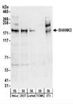
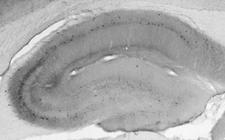
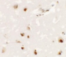
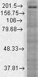
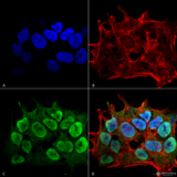
SHANK3
SH3 and multiple ankyrin repeat domains 3
Major scaffold postsynaptic density protein which interacts with multiple proteins and complexes to orchestrate the dendritic spine and synapse formation, maturation and maintenance. Interconnects receptors of the postsynaptic membrane including NMDA-type and metabotropic glutamate receptors via complexes with GKAP/PSD-95 and HOMER, respectively, and the actin-based cytoskeleton. Plays a role in the structural and functional organization of the dendritic spine and synaptic junction through the interaction with Arp2/3 and WAVE1 complex as well as the promotion of the F-actin clusters. By way of this control of actin dynamics, participates in the regulation of developing neurons growth cone motility and the NMDA receptor-signaling. Also modulates GRIA1 exocytosis and GRM5/MGLUR5 expression and signaling to control the AMPA and metabotropic glutamate receptor-mediated synaptic transmission and plasticity. May be required at an early stage of synapse formation and be inhibited by IGF1 to promote synapse maturation.
| Gene Name: | SH3 and multiple ankyrin repeat domains 3 |
| Synonyms: | SHANK3, DEL22q13.3, KIAA1650, SPANK-2, PROSAP2, PSAP2, SCZD15 |
| Target Sequences: | NM_001080420 NP_001073889.1 Q9BYB0 |
Publications (1)


If you do not find the reagent or information you require, please contact Customer.Support@LSBio.com to inquire about additional products in development.


