order histories, retained contact details for faster checkout, review submissions, and special promotions.
Forgot password?
order histories, retained contact details for faster checkout, review submissions, and special promotions.
Locations
Orders Processing,
Shipping & Receiving,
Warehouse
2 Shaker Rd Suites
B001/B101
Shirley, MA 01464
Production Lab
Floor 6, Suite 620
20700 44th Avenue W
Lynnwood, WA 98036
Telephone Numbers
Tel: +1 (206) 374-1102
Fax: +1 (206) 577-4565
Contact Us
Additional Contact Details
order histories, retained contact details for faster checkout, review submissions, and special promotions.
Forgot password?
order histories, retained contact details for faster checkout, review submissions, and special promotions.
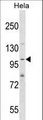
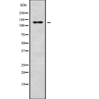
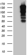
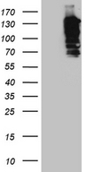
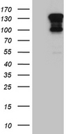
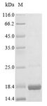
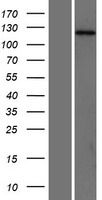
SH3PXD2A / TKS5
SH3 and PX domains 2A
Adapter protein involved in invadopodia and podosome formation, extracellular matrix degradation and invasiveness of some cancer cells. Binds matrix metalloproteinases (ADAMs), NADPH oxidases (NOXs) and phosphoinositides. Acts as an organizer protein that allows NOX1- or NOX3-dependent reactive oxygen species (ROS) generation and ROS localization. In association with ADAM12, mediates the neurotoxic effect of beta-amyloid peptide.
| Gene Name: | SH3 and PX domains 2A |
| Synonyms: | SH3PXD2A, Adapter protein TKS5, Adaptor protein TKS5, FISH, KIAA0418, SH3 and PX domains 2A, SH3 multiple domains protein 1, TSK5, SH3 multiple domains 1, SH3MD1, TKS5 |
| Target Sequences: | AX695698 IPI00023692.1 Q5TCZ1 |
Publications (2)


If you do not find the reagent or information you require, please contact Customer.Support@LSBio.com to inquire about additional products in development.


