Login
Registration enables users to use special features of this website, such as past
order histories, retained contact details for faster checkout, review submissions, and special promotions.
order histories, retained contact details for faster checkout, review submissions, and special promotions.
Forgot password?
Registration enables users to use special features of this website, such as past
order histories, retained contact details for faster checkout, review submissions, and special promotions.
order histories, retained contact details for faster checkout, review submissions, and special promotions.
Quick Order
Products
Antibodies
ELISA and Assay Kits
Research Areas
Infectious Disease
Resources
Purchasing
Reference Material
Contact Us
Locations
Orders Processing,
Shipping & Receiving,
Warehouse
2 Shaker Rd Suites
B001/B101
Shirley, MA 01464
Production Lab
Floor 6, Suite 620
20700 44th Avenue W
Lynnwood, WA 98036
Telephone Numbers
Tel: +1 (206) 374-1102
Fax: +1 (206) 577-4565
Contact Us
Additional Contact Details
Login
Registration enables users to use special features of this website, such as past
order histories, retained contact details for faster checkout, review submissions, and special promotions.
order histories, retained contact details for faster checkout, review submissions, and special promotions.
Forgot password?
Registration enables users to use special features of this website, such as past
order histories, retained contact details for faster checkout, review submissions, and special promotions.
order histories, retained contact details for faster checkout, review submissions, and special promotions.
Quick Order
| Catalog Number | Size | Price |
|---|---|---|
| LS-B10172-0.05 | 0.05 ml | $460 |
1 of 8
2 of 8
3 of 8
4 of 8
5 of 8
6 of 8
7 of 8
8 of 8
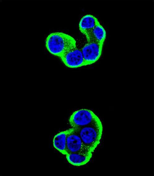
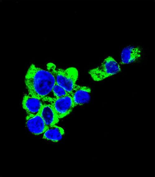
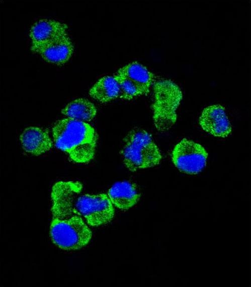
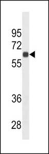
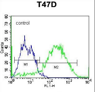
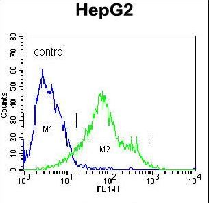
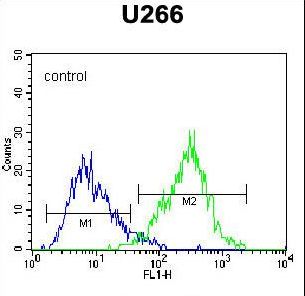
IHC‑plus™ Monoclonal Mouse anti‑Human SDC1 / Syndecan 1 / CD138 Antibody (aa210‑238, IHC, IF, WB) LS‑B10172
IHC‑plus™ Monoclonal Mouse anti‑Human SDC1 / Syndecan 1 / CD138 Antibody (aa210‑238, IHC, IF, WB) LS‑B10172

Note: This antibody replaces LS-C159300
Antibody:
SDC1 / Syndecan 1 / CD138 Mouse anti-Human Monoclonal (aa210-238) Antibody
Application:
IHC, IHC-P, IF, WB, Flo
Reactivity:
Human
Format:
Unconjugated, Unmodified
Toll Free North America
 206-374-1102
For Research Use Only
Overview
Antibody:
SDC1 / Syndecan 1 / CD138 Mouse anti-Human Monoclonal (aa210-238) Antibody
Application:
IHC, IHC-P, IF, WB, Flo
Reactivity:
Human
Format:
Unconjugated, Unmodified
Specifications
Description
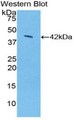
CD138 antibody LS-B10172 is an unconjugated mouse monoclonal antibody to human CD138 (SDC1 / Syndecan 1) (aa210-238). Validated for Flow, IF, IHC and WB. Tested on 20 paraffin-embedded human tissues.
Target
Human SDC1 / Syndecan 1 / CD138
Synonyms
SDC1 | CD138 antigen | SDC | SYND1 | Syndecan | Syndecan 1 | Syndecan-1 | CD138 | Syndecan proteoglycan 1
Host
Mouse
Reactivity
Human
(tested or 100% immunogen sequence identity)
Clonality
IgM
Monoclonal
Conjugations
Unconjugated
Purification
Ascites
Modifications
Unmodified
Epitope
aa210-238
Specificity
This CD138 antibody is generated from mice immunized with a KLH conjugated synthetic peptide between 210-238 amino acids from the C-terminal region of human CD138.
Applications
- IHC
- IHC - Paraffin (1:50)
- Immunofluorescence (1:10 - 1:50)
- Western blot (1:500 - 1:8000)
- Flow Cytometry (1:10 - 1:50)

|
Performing IHC? See our complete line of Immunohistochemistry Reagents including antigen retrieval solutions, blocking agents
ABC Detection Kits and polymers, biotinylated secondary antibodies, substrates and more.
|
Presentation
0.09% Sodium Azide
Storage
Maintain refrigerated at 2°C to 8°C for up to 6 months. For long term storage store at -20°C.
Restrictions
For research use only. Intended for use by laboratory professionals.
About SDC1 / Syndecan 1 / CD138
Validation
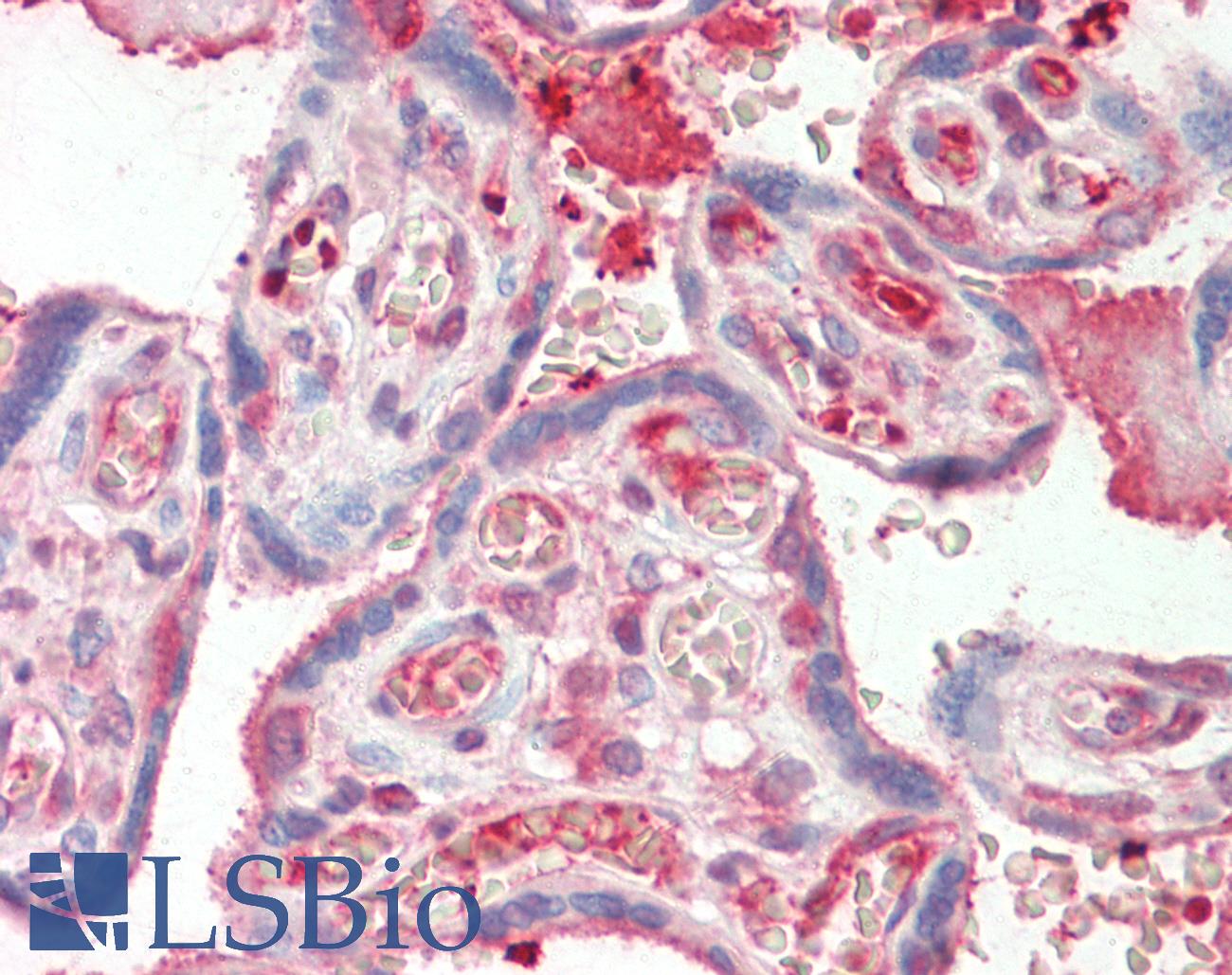
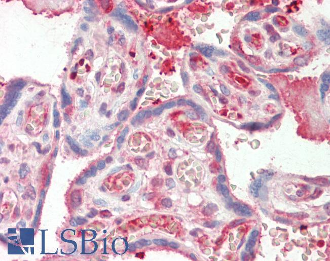
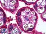
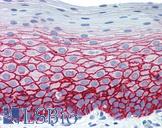
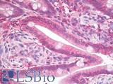
Anti-SDC1 / Syndecan 1 / CD138 antibody IHC staining of human placenta. Immunohistochemistry of formalin-fixed, paraffin-embedded tissue after heat-induced antigen retrieval. Antibody dilution 1:50.
Anti-SDC1 / Syndecan 1 / CD138 antibody IHC staining of human placenta. Immunohistochemistry of formalin-fixed, paraffin-embedded tissue after heat-induced antigen retrieval. Antibody dilution 1:50.
See More About...
LSBio Ratings
IHC-plus™ SDC1 / Syndecan 1 / CD138 Antibody (aa210-238) for IHC, IF/Immunofluorescence, WB/Western, Flow LS-B10172 has an LSBio Rating of
Laboratory Validation Score (4)
Learn more about The LSBio Ratings Algorithm
Publications (0)
Customer Reviews (0)
Featured Products
Species:
Human, Mouse
Applications:
IHC, IHC - Paraffin, Western blot
Species:
Human
Applications:
IHC, IHC - Paraffin, IHC - Frozen, Immunofluorescence, Western blot, Flow Cytometry
Species:
Human
Applications:
Western blot, ELISA
Species:
Human
Applications:
IHC, Flow Cytometry, ELISA
Species:
Rat
Applications:
Western blot
Species:
Human, Mouse, Rat
Applications:
IHC, IHC - Paraffin, Western blot, ELISA
Request SDS/MSDS
To request an SDS/MSDS form for this product, please contact our Technical Support department at:
Technical.Support@LSBio.com
Requested From: United States
Date Requested: 4/19/2024
Date Requested: 4/19/2024


