Login
Registration enables users to use special features of this website, such as past
order histories, retained contact details for faster checkout, review submissions, and special promotions.
order histories, retained contact details for faster checkout, review submissions, and special promotions.
Forgot password?
Registration enables users to use special features of this website, such as past
order histories, retained contact details for faster checkout, review submissions, and special promotions.
order histories, retained contact details for faster checkout, review submissions, and special promotions.
Quick Order
Products
Antibodies
ELISA and Assay Kits
Research Areas
Infectious Disease
Resources
Purchasing
Reference Material
Contact Us
Locations
Orders Processing,
Shipping & Receiving,
Warehouse
2 Shaker Rd Suites
B001/B101
Shirley, MA 01464
Production Lab
Floor 6, Suite 620
20700 44th Avenue W
Lynnwood, WA 98036
Telephone Numbers
Tel: +1 (206) 374-1102
Fax: +1 (206) 577-4565
Contact Us
Additional Contact Details
Login
Registration enables users to use special features of this website, such as past
order histories, retained contact details for faster checkout, review submissions, and special promotions.
order histories, retained contact details for faster checkout, review submissions, and special promotions.
Forgot password?
Registration enables users to use special features of this website, such as past
order histories, retained contact details for faster checkout, review submissions, and special promotions.
order histories, retained contact details for faster checkout, review submissions, and special promotions.
Quick Order
| Catalog Number | Size | Price |
|---|---|---|
| LS-C60076-100 | 100 µg (1.05 mg/ml) | $435 |
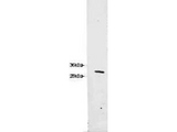
![RFP / Red Fluorescent Protein Antibody - Anti-RFP Antibody - Western Blot. Western blot of RFP recombinant protein detected with polyclonal anti-RFP antibody. Lane 1 shows no reaction against a GFP recombinant protein present in 10 ug of HeLa cell extract. Lane 2 shows a single band detected in 10 ug of a HeLa lysate containing RFP recombinant protein as a 27 kD band. A 4-12% Bis-Tris gradient gel (Invitrogen) was used for SDS-PAGE. The membrane was blocked and then probed with Anti-RFP diluted 1:2500 for 1 h at RT followed by washes and reaction with a 1:5000 dilution of IRDye800 conjugated Goat-a-Rabbit IgG [H&L] MX (. IRDye800 fluorescence image was captured using the Odyssey Infrared Imaging System developed by LI-COR. IRDye is a trademark of LI-COR, Inc. Other detection systems will yield similar results.](https://lsbio-7d62.kxcdn.com/image2/rfp-red-fluorescent-protein-antibody-ls-c60076/62809_194421.jpg)
1 of 2
2 of 2
Polyclonal Rabbit anti‑Discosoma RFP / Red Fluorescent Protein Antibody (IHC, IF, WB) LS‑C60076
Polyclonal Rabbit anti‑Discosoma RFP / Red Fluorescent Protein Antibody (IHC, IF, WB) LS‑C60076

Antibody:
RFP / Red Fluorescent Protein Rabbit anti-Discosoma Polyclonal Antibody
Application:
IHC, IF, WB, Flo, ELISA
Reactivity:
Discosoma
Format:
Unconjugated, Unmodified
Toll Free North America
 206-374-1102
For Research Use Only
Overview
Antibody:
RFP / Red Fluorescent Protein Rabbit anti-Discosoma Polyclonal Antibody
Application:
IHC, IF, WB, Flo, ELISA
Reactivity:
Discosoma
Format:
Unconjugated, Unmodified
Specifications
Description
Red Fluorescent Protein antibody LS-C60076 is an unconjugated rabbit polyclonal antibody to discosoma Red Fluorescent Protein (RFP). Validated for ELISA, Flow, IF, IHC and WB. Cited in 1 publication.
Host
Rabbit
Reactivity
Discosoma
(tested or 100% immunogen sequence identity)
Clonality
IgG
Polyclonal
Conjugations
Unconjugated
Purification
Affinity chromatography
Modifications
Unmodified
Immunogen
The immunogen is a Red Fluorescent Protein (RFP) fusion protein corresponding to the full length amino acid sequence (234aa) derived from the mushroom polyp coral Discosoma.
Specificity
Assay by immunoelectrophoresis resulted in a single precipitin arc against anti-Rabbit Serum and purified and partially purified Red Fluorescent Protein (Discosoma). No reaction was observed against Human, Mouse or Rat serum proteins.
Applications
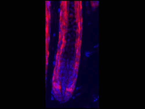
- IHC (1:200 - 1:2000)
- Immunofluorescence (1:200 - 1:2000)
- Western blot (1:1000 - 1:5000)
- Flow Cytometry (1:200 - 1:2000)
- ELISA (1:28700 - 1:48700)

|
Performing IHC? See our complete line of Immunohistochemistry Reagents including antigen retrieval solutions, blocking agents
ABC Detection Kits and polymers, biotinylated secondary antibodies, substrates and more.
|
Usage
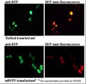
Polyclonal anti-RFP is designed to detect RFP and its variants. This antibody can be used to detect RFP by ELISA (sandwich or capture) for the direct binding of antigen. Biotin conjugated polyclonal anti-RFP used in a sandwich ELISA with unconjugated anti-RFP is well suited to titrate RFP in solution. Fluorochrome conjugated polyclonal anti-RFP can be used to detect RFP by immunofluorescence microscopy in cell expression systems and can detect RFP containing inserts. Significant amplification of signal is achieved using fluorochrome conjugated polyclonal anti-RFP relative to the fluorescence of RFP alone. For immunoblotting use either alkaline phosphatase or peroxidase conjugated polyclonal anti-RFP to detect RFP or RFP containing proteins on western blots. Optimal titers for applications should be determined by the researcher.
Presentation
0.02 M Potassium Phosphate, pH 7.2, 0.15 M NaCl, 0.01% Sodium Azide
Storage
Store vial at -20°C prior to opening. This product is stable for several weeks at 4°C as an undiluted liquid. Dilute only prior to immediate use. For extended storage aliquot contents and freeze at -20°C or below. Avoid freeze-thaw cycles.
Restrictions
For research use only. Intended for use by laboratory professionals.
LSBio Ratings
RFP / Red Fluorescent Protein Antibody for IHC, IF/Immunofluorescence, WB/Western, Flow, ELISA LS-C60076 has an LSBio Rating of
Publications (4)
Learn more about The LSBio Ratings Algorithm
Publications (1)
Increased cell-intrinsic excitability induces synaptic changes in new neurons in the adult dentate gyrus that require Npas4. Sim S, Antolin S, Lin CW, Lin Y, Lin YX, Lois C. The Journal of neuroscience : the official journal of the Society for Neuroscience. 2013 May;33:7928-40.
Customer Reviews (0)
Featured Products
Species:
Discosoma
Applications:
Western blot, Immunoprecipitation, ELISA
Species:
Applications:
ICC, Western blot
![RFP / Red Fluorescent Protein Antibody - Western blot of RFP recombinant protein detected with polyclonal anti-RFP antibody. Lane 1 shows no reaction against a GFP recombinant protein present in 10 ug of HeLa cell extract. Lane 2 shows a single band detected in 10 ug of a HeLa lysate containing RFP recombinant protein. polyclonal anti-RFP detects a 27 kDa band corresponding to the epitope tag RFP. A 4-12% Bis-Tris gradient gel (Invitrogen) was used for SDS-PAGE. The protein was transferred to nitrocellulose using standard methods. After blocking the membrane was probed with the primary antibody diluted 1:2,500 for 1 h at room temperature followed by washes and reaction with a 1:5,000 dilution of IRDye™800 conjugated Goat-a-Rabbit IgG [H&L] MX. IRDye™800 fluorescence image was captured using the Odyssey® Infrared Imaging System developed by LI-COR. IRDye is a trademark of LI-COR, Inc. Other detection systems will yield similar results.](https://lsbio-7d62.kxcdn.com//image2/rfp-red-fluorescent-protein-antibody-fitc-ls-c154267/590804_5351757.png)
Species:
Discosoma
Applications:
Immunofluorescence, Western blot, Fluorophore-Linked Immunosorbent Assay
Species:
Discosoma
Applications:
Western blot, ELISA
Reactivity:
Pig
Range:
0.156-10 ng/ml
Request SDS/MSDS
To request an SDS/MSDS form for this product, please contact our Technical Support department at:
Technical.Support@LSBio.com
Requested From: United States
Date Requested: 4/18/2024
Date Requested: 4/18/2024


