order histories, retained contact details for faster checkout, review submissions, and special promotions.
Forgot password?
order histories, retained contact details for faster checkout, review submissions, and special promotions.
Locations
Orders Processing,
Shipping & Receiving,
Warehouse
2 Shaker Rd Suites
B001/B101
Shirley, MA 01464
Production Lab
Floor 6, Suite 620
20700 44th Avenue W
Lynnwood, WA 98036
Telephone Numbers
Tel: +1 (206) 374-1102
Fax: +1 (206) 577-4565
Contact Us
Additional Contact Details
order histories, retained contact details for faster checkout, review submissions, and special promotions.
Forgot password?
order histories, retained contact details for faster checkout, review submissions, and special promotions.
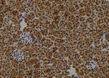
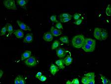
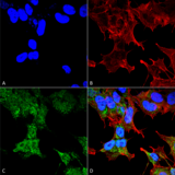
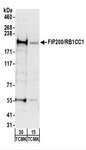
RB1CC1 / CC1
RB1-inducible coiled-coil 1
Plays a role as a modulator of TGF-beta-signaling by restricting substrate specificity of RNF111. Involved in autophagy. Regulates early events but also late events of autophagosome formation through direct interaction with Atg16L1. Required for the formation of the autophagosome-like double-membrane structure that surrounds the Salmonella-containing vacuole (SCV) duting S.typhimurium infection and subsequent xenophagy. Autophagy positively regulates repair of DNA damage induced by ionizing radiation and negatively regulates apoptosis. Plays an indispensible role in fetal hematopoiesis and in the regulation of neuronal homeostasis (By similarity). Implicated in the regulation of RB1 expression. Functions as a DNA-binding transcription factor. Is a potent regulator of the RB1 pathway and a mediator that plays a crucial role in muscular differentiation. Expression is, thus, a prerequisite for myogenic differentiation. Inhibits PTK2/FAK1 and PTK2B/PYK2 activity and activation of downstream signaling pathways.
| Gene Name: | RB1-inducible coiled-coil 1 |
| Synonyms: | RB1CC1, CC1, DRAGOU14, FIP200, RBICC, KIAA0203, RB1-inducible coiled-coil 1 |
| Target Sequences: | NM_014781 NP_055596.3 Q8TDY2 |


If you do not find the reagent or information you require, please contact Customer.Support@LSBio.com to inquire about additional products in development.


