order histories, retained contact details for faster checkout, review submissions, and special promotions.
Forgot password?
order histories, retained contact details for faster checkout, review submissions, and special promotions.
Locations
Orders Processing,
Shipping & Receiving,
Warehouse
2 Shaker Rd Suites
B001/B101
Shirley, MA 01464
Production Lab
Floor 6, Suite 620
20700 44th Avenue W
Lynnwood, WA 98036
Telephone Numbers
Tel: +1 (206) 374-1102
Fax: +1 (206) 577-4565
Contact Us
Additional Contact Details
order histories, retained contact details for faster checkout, review submissions, and special promotions.
Forgot password?
order histories, retained contact details for faster checkout, review submissions, and special promotions.
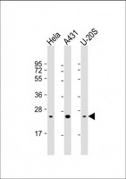
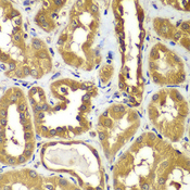
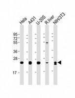
RAB1B
RAB1B, member RAS oncogene family
The small GTPases Rab are key regulators of intracellular membrane trafficking, from the formation of transport vesicles to their fusion with membranes. Rabs cycle between an inactive GDP-bound form and an active GTP-bound form that is able to recruit to membranes different set of downstream effectors directly responsible for vesicle formation, movement, tethering and fusion. RAB1B regulates vesicular transport between the endoplasmic reticulum and successive Golgi compartments. Plays a role in the initial events of the autophagic vacuole development which take place at specialized regions of the endoplasmic reticulum.
| Gene Name: | RAB1B, member RAS oncogene family |
| Family/Subfamily: | Ras GTPase superfamily IPR001806 , RAS oncogene |
| Synonyms: | RAB1B, Ras-related protein Rab-1B, Small GTP-binding protein |
| Target Sequences: | NM_030981 NP_112243.1 Q9H0U4 |

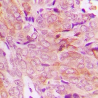
![RAB1B Antibody - Immunoperoxidase of monoclonal antibody to RAB1B on formalin-fixed paraffin-embedded human endometrium. [antibody concentration 3 ug/ml]](https://lsbio-7d62.kxcdn.com/image2/rab1b-antibody-clone-1b2-ls-c134105/53758_5176318.jpg)


If you do not find the reagent or information you require, please contact Customer.Support@LSBio.com to inquire about additional products in development.


