Login
Registration enables users to use special features of this website, such as past
order histories, retained contact details for faster checkout, review submissions, and special promotions.
order histories, retained contact details for faster checkout, review submissions, and special promotions.
Forgot password?
Registration enables users to use special features of this website, such as past
order histories, retained contact details for faster checkout, review submissions, and special promotions.
order histories, retained contact details for faster checkout, review submissions, and special promotions.
Quick Order
Products
Antibodies
ELISA and Assay Kits
Research Areas
Infectious Disease
Resources
Purchasing
Reference Material
Contact Us
Locations
Orders Processing,
Shipping & Receiving,
Warehouse
2 Shaker Rd Suites
B001/B101
Shirley, MA 01464
Production Lab
Floor 6, Suite 620
20700 44th Avenue W
Lynnwood, WA 98036
Telephone Numbers
Tel: +1 (206) 374-1102
Fax: +1 (206) 577-4565
Contact Us
Additional Contact Details
Login
Registration enables users to use special features of this website, such as past
order histories, retained contact details for faster checkout, review submissions, and special promotions.
order histories, retained contact details for faster checkout, review submissions, and special promotions.
Forgot password?
Registration enables users to use special features of this website, such as past
order histories, retained contact details for faster checkout, review submissions, and special promotions.
order histories, retained contact details for faster checkout, review submissions, and special promotions.
Quick Order
| Catalog Number | Size | Price |
|---|---|---|
| LS-B11047-50 | 50 µl | $460 |

1 of 3
2 of 3
3 of 3
IHC‑plus™ Polyclonal Rabbit anti‑Human NUMA1 / NUMA Antibody (aa900‑950, IHC, IF) LS‑B11047
IHC‑plus™ Polyclonal Rabbit anti‑Human NUMA1 / NUMA Antibody (aa900‑950, IHC, IF) LS‑B11047

Note: This antibody replaces LS-C286543
Antibody:
NUMA1 / NUMA Rabbit anti-Human Polyclonal (aa900-950) Antibody
Application:
IHC, IHC-P, IF
Reactivity:
Human
Format:
Unconjugated, Unmodified
Toll Free North America
 206-374-1102
For Research Use Only
Overview
Antibody:
NUMA1 / NUMA Rabbit anti-Human Polyclonal (aa900-950) Antibody
Application:
IHC, IHC-P, IF
Reactivity:
Human
Format:
Unconjugated, Unmodified
Specifications
Description
NUMA antibody LS-B11047 is an unconjugated rabbit polyclonal antibody to human NUMA (NUMA1) (aa900-950). Validated for IF and IHC. Tested on 20 paraffin-embedded human tissues. Cited in 3 publications.
Target
Human NUMA1 / NUMA
Synonyms
NUMA1 | Structural nuclear protein | NuMA protein | SP-H antigen | NUMA
Host
Rabbit
Reactivity
Human
(tested or 100% immunogen sequence identity)
Predicted
Chimpanzee, Gorilla (at least 90% immunogen sequence identity)
Clonality
IgG
Polyclonal
Conjugations
Unconjugated
Purification
Affinity purified
Modifications
Unmodified
Immunogen
A portion of GeneID: 4926 corresponding to amino acids between 900 and 950 of UniprotID: Q14980
Epitope
aa900-950
Specificity
Region between residue 900 and 950 of human nuclear mitotic apparatus protein 1 using the numbering given in entry NP_006176.2 (GeneID 4926).
Applications
- IHC
- IHC - Paraffin (1:100)
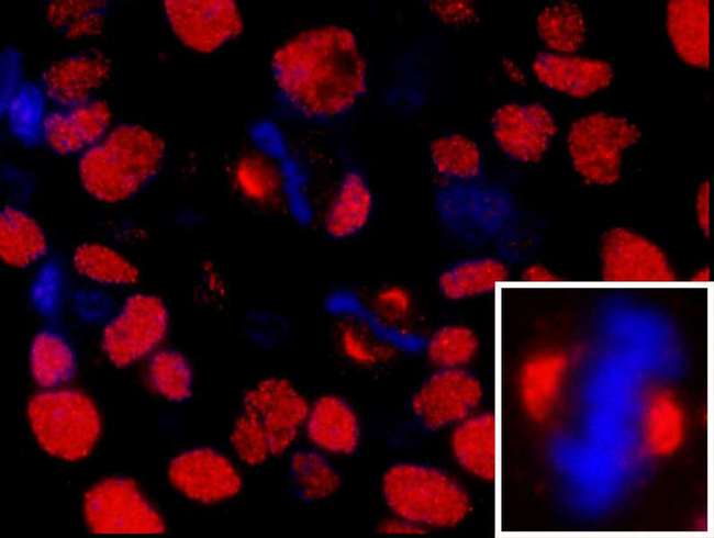
- Immunofluorescence (1:50 - 1:500)

|
Performing IHC? See our complete line of Immunohistochemistry Reagents including antigen retrieval solutions, blocking agents
ABC Detection Kits and polymers, biotinylated secondary antibodies, substrates and more.
|
Usage
IHC-IF: 1:50-1:500
Manufacturer
Bethyl Laboratories, Inc.
Presentation
TBS, 0.09% Sodium Azide, 0.1% BSA
Storage
Store at 2-8°C for up to one year.
Restrictions
For research use only. Intended for use by laboratory professionals.
About NUMA1 / NUMA
Validation
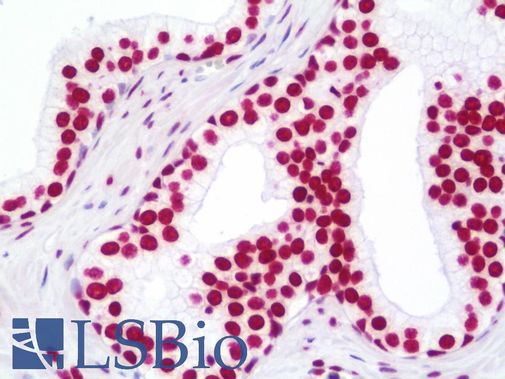
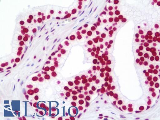
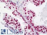
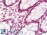
Anti-NUMA1 / NUMA antibody IHC staining of human prostate. Immunohistochemistry of formalin-fixed, paraffin-embedded tissue after heat-induced antigen retrieval.
Anti-NUMA1 / NUMA antibody IHC staining of human prostate. Immunohistochemistry of formalin-fixed, paraffin-embedded tissue after heat-induced antigen retrieval.
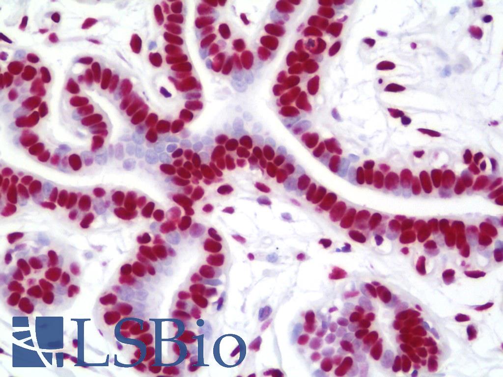
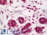
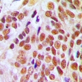
Anti-NUMA1 / NUMA antibody IHC staining of human breast. Immunohistochemistry of formalin-fixed, paraffin-embedded tissue after heat-induced antigen retrieval. Antibody dilution 1:100.
Anti-NUMA1 / NUMA antibody IHC staining of human breast. Immunohistochemistry of formalin-fixed, paraffin-embedded tissue after heat-induced antigen retrieval. Antibody dilution 1:100.
See More About...
LSBio Ratings
IHC-plus™ NUMA1 / NUMA Antibody (aa900-950) for IHC, IF/Immunofluorescence LS-B11047 has an LSBio Rating of
Publications (4.2)
Laboratory Validation Score (4)
Learn more about The LSBio Ratings Algorithm
Publications (3)
Spectrum of Posttransplant Lymphoproliferations in NSG Mice and Their Association With EBV Infection After Engraftment of Pediatric Solid Tumors. Heather Tillman, Peter Vogel, Tiffani Rogers, Walter Akers, Jerold E Rehg. Veterinary pathology. 2020 May;57:445-456.
Morphologic and Immunohistochemical Characterization of Spontaneous Lymphoma/Leukemia in NSG Mice. Heather Tillman, Laura J Janke, Amy Funk, Peter Vogel, Jerold E Rehg. Veterinary pathology. 2020 January;57:160-171.
Development of Mast Cell and Eosinophil Hyperplasia and HLH/MAS-Like Disease in NSG-SGM3 Mice Receiving Human CD34+ Hematopoietic Stem Cells or Patient-Derived Leukemia Xenografts. Laura J Janke, Denise M Imai, Heather Tillman, Rosalinda Doty, Mark J Hoenerhoff, Jiajie J Xu, Zachary T Freeman, Portia Allen, Natalie Wall Fowlkes, Ilaria Iacobucci, Kirsten Dickerson, Charles G Mullighan, Peter Vogel, Jerold E Rehg. Veterinary pathology. 2021 January;58:181-204.
Customer Reviews (0)
Featured Products
Species:
Human
Applications:
IHC, IHC - Paraffin, Western blot, Immunoprecipitation, ELISA
Species:
Human
Applications:
IHC, IHC - Paraffin, ICC, Western blot

Species:
Human
Applications:
IHC, IHC - Paraffin, Western blot, ELISA
Species:
Human
Applications:
IHC, IHC - Paraffin, Western blot, Peptide Enzyme-Linked Immunosorbent Assay
Species:
Human
Applications:
IHC, IHC - Paraffin, Western blot
Request SDS/MSDS
To request an SDS/MSDS form for this product, please contact our Technical Support department at:
Technical.Support@LSBio.com
Requested From: United States
Date Requested: 4/18/2024
Date Requested: 4/18/2024


