Login
Registration enables users to use special features of this website, such as past
order histories, retained contact details for faster checkout, review submissions, and special promotions.
order histories, retained contact details for faster checkout, review submissions, and special promotions.
Forgot password?
Registration enables users to use special features of this website, such as past
order histories, retained contact details for faster checkout, review submissions, and special promotions.
order histories, retained contact details for faster checkout, review submissions, and special promotions.
Quick Order
Products
Antibodies
ELISA and Assay Kits
Research Areas
Infectious Disease
Resources
Purchasing
Reference Material
Contact Us
Locations
Orders Processing,
Shipping & Receiving,
Warehouse
2 Shaker Rd Suites
B001/B101
Shirley, MA 01464
Production Lab
Floor 6, Suite 620
20700 44th Avenue W
Lynnwood, WA 98036
Telephone Numbers
Tel: +1 (206) 374-1102
Fax: +1 (206) 577-4565
Contact Us
Additional Contact Details
Login
Registration enables users to use special features of this website, such as past
order histories, retained contact details for faster checkout, review submissions, and special promotions.
order histories, retained contact details for faster checkout, review submissions, and special promotions.
Forgot password?
Registration enables users to use special features of this website, such as past
order histories, retained contact details for faster checkout, review submissions, and special promotions.
order histories, retained contact details for faster checkout, review submissions, and special promotions.
Quick Order
| Catalog Number | Size | Price |
|---|---|---|
| LS-C312448-10 | 10 µg | $318 |
| LS-C312448-100 | 100 µg (0.1 mg/ml) | $470 |
1 of 2
2 of 2
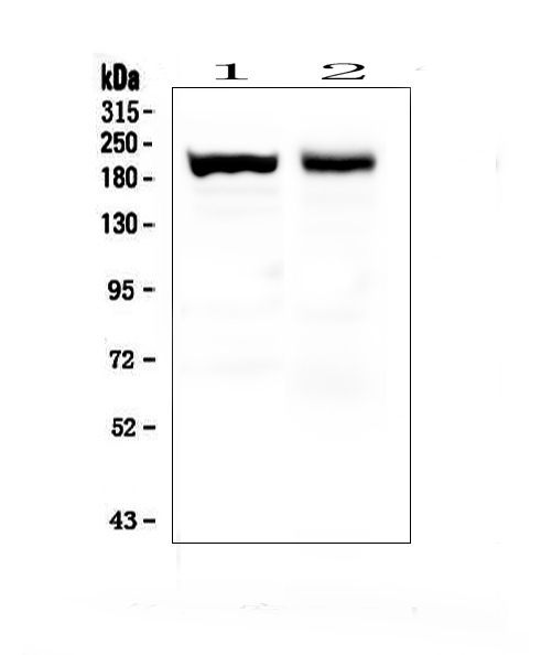
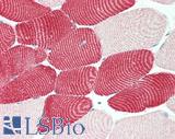
Monoclonal Mouse anti‑Human MYH7 Antibody (clone IML‑64, IHC, WB) LS‑C312448
Monoclonal Mouse anti‑Human MYH7 Antibody (clone IML‑64, IHC, WB) LS‑C312448

Antibody:
MYH7 Mouse anti-Human Monoclonal (IML-64) Antibody
Application:
IHC, IHC-P, WB
Reactivity:
Human, Mouse, Rat, Rabbit
Format:
Unconjugated, Unmodified
Toll Free North America
 206-374-1102
For Research Use Only
Overview
Antibody:
MYH7 Mouse anti-Human Monoclonal (IML-64) Antibody
Application:
IHC, IHC-P, WB
Reactivity:
Human, Mouse, Rat, Rabbit
Format:
Unconjugated, Unmodified
Specifications
Description
MYH7 antibody LS-C312448 is an unconjugated mouse monoclonal antibody to MYH7 from human. It is reactive with human, mouse, rat and other species. Validated for IHC and WB. Cited in 2 publications.
Target
Human MYH7
Synonyms
MYH7 | Beta-myosin heavy chain | MYHCB | Myosin heavy chain (AA 1-96) | MPD1 | Myhc-slow | Myopathy, distal 1 | Myosin heavy chain 7 | MyHC-beta | Myosin-7 | CMD1S | CMH1 | SPMD | SPMM
Host
Mouse
Reactivity
Human, Mouse, Rat, Rabbit
(tested or 100% immunogen sequence identity)
Clonality
IgG1
Monoclonal
Clone
IML-64
Conjugations
Unconjugated
Purification
Ascites
Modifications
Unmodified
Immunogen
Human skeletal muscle myosin purified from myofibrils.
Specificity
MYH7: Both wild type and variant Gln-403 aredetected in skeletal muscle (at protein level).
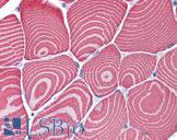
Applications
- IHC
- IHC - Paraffin
- Western blot

|
Performing IHC? See our complete line of Immunohistochemistry Reagents including antigen retrieval solutions, blocking agents
ABC Detection Kits and polymers, biotinylated secondary antibodies, substrates and more.
|
Presentation
Lyophilized from ascites, 1.2% sodium acetate, 2mg BSA, 0.01mg sodium azide.
Reconstitution
Add 1ml of PBS buffer will yield a concentration of 100ug/ml.
Storage
At -20°C for 1 year. After reconstitution, at 4°C for 1 month. It can also be aliquotted and stored frozen at -20°C for a longer time. Avoid freeze-thaw cycles.
Restrictions
For research use only. Intended for use by laboratory professionals.
LSBio Ratings
MYH7 Antibody (clone IML-64) for IHC, WB/Western LS-C312448 has an LSBio Rating of
Publications (4.1)
Learn more about The LSBio Ratings Algorithm
Publications (2)
Characterization of cytoskeleton features and maturation status of cultured human iPSC-derived cardiomyocytes. Christian Zuppinger, George Gibbons, Priyanka Dutta-Passecker, Adrian Segiser, Henriette Most, Thomas M Suter. European journal of histochemistry : EJH. 2017 Jun;61:2763.
3D Co-culture of hiPSC-Derived Cardiomyocytes With Cardiac Fibroblasts Improves Tissue-Like Features of Cardiac Spheroids. Philippe Beauchamp, Christopher B Jackson, Lijo Cherian Ozhathil, Irina Agarkova, Cristi L Galindo, Douglas B Sawyer, Thomas M Suter, Christian Zuppinger. Frontiers in molecular biosciences. 2020 Feb;7:14.
Customer Reviews (0)
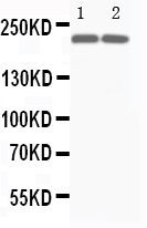
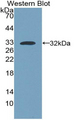
Featured Products
Species:
Mouse, Human
Applications:
IHC, IHC - Paraffin, Western blot
Species:
Human, Mouse, Rat
Applications:
IHC, IHC - Paraffin, Western blot
Request SDS/MSDS
To request an SDS/MSDS form for this product, please contact our Technical Support department at:
Technical.Support@LSBio.com
Requested From: United States
Date Requested: 4/19/2024
Date Requested: 4/19/2024


