order histories, retained contact details for faster checkout, review submissions, and special promotions.
Forgot password?
order histories, retained contact details for faster checkout, review submissions, and special promotions.
Locations
Orders Processing,
Shipping & Receiving,
Warehouse
2 Shaker Rd Suites
B001/B101
Shirley, MA 01464
Production Lab
Floor 6, Suite 620
20700 44th Avenue W
Lynnwood, WA 98036
Telephone Numbers
Tel: +1 (206) 374-1102
Fax: +1 (206) 577-4565
Contact Us
Additional Contact Details
order histories, retained contact details for faster checkout, review submissions, and special promotions.
Forgot password?
order histories, retained contact details for faster checkout, review submissions, and special promotions.
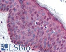
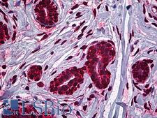
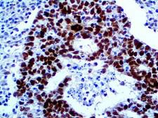
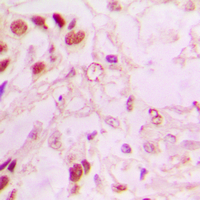
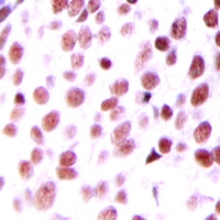
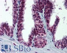
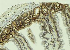
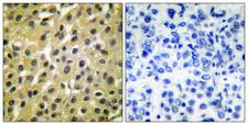
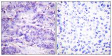
MDM2
MDM2 oncogene, E3 ubiquitin protein ligase
E3 ubiquitin-protein ligase that mediates ubiquitination of p53/TP53, leading to its degradation by the proteasome. Inhibits p53/TP53- and p73/TP73-mediated cell cycle arrest and apoptosis by binding its transcriptional activation domain. Also acts as a ubiquitin ligase E3 toward itself and ARRB1. Permits the nuclear export of p53/TP53. Promotes proteasome-dependent ubiquitin-independent degradation of retinoblastoma RB1 protein. Inhibits DAXX-mediated apoptosis by inducing its ubiquitination and degradation. Component of the TRIM28/KAP1-MDM2-p53/TP53 complex involved in stabilizing p53/TP53. Also component of the TRIM28/KAP1-ERBB4-MDM2 complex which links growth factor and DNA damage response pathways. Mediates ubiquitination and subsequent proteasome degradation of DYRK2 in nucleus. Ubiquitinates IGF1R and SNAI1 and promotes them to proteasomal degradation.
| Gene Name: | MDM2 oncogene, E3 ubiquitin protein ligase |
| Synonyms: | MDM2, ACTFS, Double minute 2 protein, Hdm2, MDM2 variant FB30, HDMX, Oncoprotein Mdm2, p53-binding protein Mdm2, MDM2 variant FB28 |
| Target Sequences: | NM_002392 NP_002383.2 Q00987 |
Publications (3)

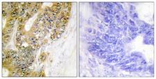

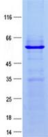
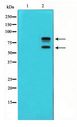
![MDM2 Antibody - Anti-MDM2 Antibody - Western Blot. Western blot using affinity purified Anti-MDM2 (Rabbit) is shown to detect a band (arrow) corresponding to mouse MDM2 protein present in mouse MEF cells (lane 2) but not human kidney HEK293 cells (lane 1). Approximately 35 ug of lysate was separated by 4-20% Tris Glycine SDS-PAGE. After blocking the membrane with 5% normal goat serum, 0.5% BLOTTO in PBS, the membrane was probed for overnight at 4° with the primary antibody diluted to 1:500 in 1% normal goat serum, 0.1% BLOTTO in PBS. The membrane was washed and reacted with a 1:10000 dilution of IRDye800 conjugated Gt-a-Rabbit IgG [H&L] ( for 45 min at room temperature (800 nm channel, green). Molecular weight estimation was made by comparison to prestained MW markers indicated at the right (700 nm channel, red). IRDye800 fluorescence image was captured using the Ddyssey Infrared Imaging System developed by LI-COR. IRDye is a trademark of LI-COR, Inc. Other detection systems will yield similar results.](https://lsbio-7d62.kxcdn.com/image2/mdm2-antibody-ls-c19046/62938_910982.jpg)

If you do not find the reagent or information you require, please contact Customer.Support@LSBio.com to inquire about additional products in development.


