Login
Registration enables users to use special features of this website, such as past
order histories, retained contact details for faster checkout, review submissions, and special promotions.
order histories, retained contact details for faster checkout, review submissions, and special promotions.
Forgot password?
Registration enables users to use special features of this website, such as past
order histories, retained contact details for faster checkout, review submissions, and special promotions.
order histories, retained contact details for faster checkout, review submissions, and special promotions.
Quick Order
Products
Antibodies
ELISA and Assay Kits
Research Areas
Infectious Disease
Resources
Purchasing
Reference Material
Contact Us
Locations
Orders Processing,
Shipping & Receiving,
Warehouse
2 Shaker Rd Suites
B001/B101
Shirley, MA 01464
Production Lab
Floor 6, Suite 620
20700 44th Avenue W
Lynnwood, WA 98036
Telephone Numbers
Tel: +1 (206) 374-1102
Fax: +1 (206) 577-4565
Contact Us
Additional Contact Details
Login
Registration enables users to use special features of this website, such as past
order histories, retained contact details for faster checkout, review submissions, and special promotions.
order histories, retained contact details for faster checkout, review submissions, and special promotions.
Forgot password?
Registration enables users to use special features of this website, such as past
order histories, retained contact details for faster checkout, review submissions, and special promotions.
order histories, retained contact details for faster checkout, review submissions, and special promotions.
Quick Order
| Catalog Number | Size | Price |
|---|---|---|
| LS-B391-100 | 100 µg (0.98 mg/ml) | $505 |

1 of 2
2 of 2
PathPlus™ Polyclonal Rabbit anti‑Human MCM2 Antibody (aa15‑40, IHC, WB) LS‑B391
PathPlus™ Polyclonal Rabbit anti‑Human MCM2 Antibody (aa15‑40, IHC, WB) LS‑B391

Note: This antibody replaces LS-C18788
Antibody:
MCM2 Rabbit anti-Human Polyclonal (aa15-40) Antibody
Application:
IHC, IHC-P, WB, ELISA
Reactivity:
Human, Mouse, Rat, S. cerevisiae
Format:
Unconjugated, Unmodified
Toll Free North America
 206-374-1102
For Research Use Only
Overview
Antibody:
MCM2 Rabbit anti-Human Polyclonal (aa15-40) Antibody
Application:
IHC, IHC-P, WB, ELISA
Reactivity:
Human, Mouse, Rat, S. cerevisiae
Format:
Unconjugated, Unmodified
Specifications
Description
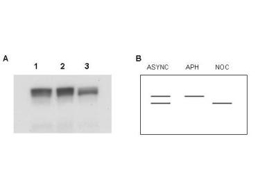
MCM2 is a component of the MCM complex, a replicative helicase essential for 'once per cell cycle' DNA replication initiation and elongation in eukaryotic cells. MCM2 is required for entry into the S phase and for cell division. MCM2 is a useful marker in immunohistochemistry for characterizing a number of cancers. It is overexpressed in precancerous and cancerous cervical lesions and upregulation is associated with high-risk type HPV. In squamous cell and adenocarcinoma of the gallbladder, MCM2 positivity is associated with metastasis and poor prognosis. It has similar prognostic value in oral dysplasia and ovarian serous neoplasms, where expression increases with severity of tumor grade. In immunohistochemistry of normal tissue, MCM2 has nuclear positivity in proliferative cells throughout the body.
References: Int J Clin Exp Pathol. 2015; 8(1): 875–880, PMID: 25755789; Mol Med Rep. 2016 Nov; 14(5): 4581–4592, PMID: 27748889; Applied Immunohistochemistry & Molecular Morphology. 2018 Aug. 26(7):509–513, DOI: 10.1097/PAI.0000000000000330; British Journal of Cancer. 2004. 90:1583–1590, PMID: 15083189
Target
Human MCM2
Synonyms
MCM2 | BM28 | Cell devision cycle-like 1 | D3S3194 | Cdc19 | Cyclin-like 1 | KIAA0030 | Nuclear protein BM28 | CDCL1 | MITOTIN
Host
Rabbit
Reactivity
Human, Mouse, Rat, S. cerevisiae
(tested or 100% immunogen sequence identity)
Predicted
Human, Mouse, Rat, S. cerevisiae (at least 90% immunogen sequence identity)
Clonality
IgG
Polyclonal
Conjugations
Unconjugated
Purification
Affinity purified
Modifications
Unmodified
Immunogen
This affinity purified antibody was prepared from whole rabbit serum produced by repeated immunizations with a synthetic peptide corresponding to an N-Terminal region near amino acids 15-40 of human MCM2 protein.
Epitope
aa15-40
Specificity
Approximately equivalent reactivity occurs against both unphosphorylated and phosphorylated forms of human MCM2 (phosphorylated at residues pS26 and pS27). A BLAST analysis was used to suggest cross reactivity with MCM2 proteins from human, mouse, rat and S. cerevisiae based on 100% homology with the immunizing sequence. Reactivity against homologues from other sources is not known.
Applications
- IHC
- IHC - Paraffin (5 µg/ml)
- Western blot (1:500 - 1:2000)
- ELISA (1:10000 - 1:50000)

|
Performing IHC? See our complete line of Immunohistochemistry Reagents including antigen retrieval solutions, blocking agents
ABC Detection Kits and polymers, biotinylated secondary antibodies, substrates and more.
|
Usage
Immunohistochemistry: LS-B391 was validated for use in immunohistochemistry on a panel of 21 formalin-fixed, paraffin-embedded (FFPE) human tissues after heat induced antigen retrieval in pH 6.0 citrate buffer. After incubation with the primary antibody, slides were incubated with biotinylated secondary antibody, followed by alkaline phosphatase-streptavidin and chromogen. The stained slides were evaluated by a pathologist to confirm staining specificity. The optimal working concentration for LS-B391 was determined to be 5 ug/ml.
Presentation
0.02 M Potassium Phosphate, pH 7.2, 0.15 M NaCl, 0.01% Sodium Azide
Storage
Aliquot and store at -20°C. Avoid freeze-thaw cycles.
Restrictions
For research use only. Intended for use by laboratory professionals.
About MCM2
Validation
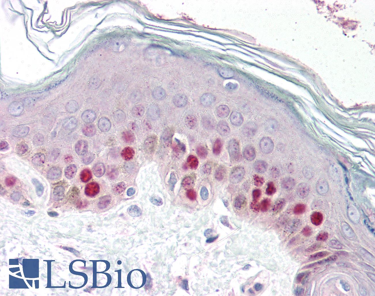
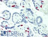
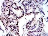
Anti-MCM2 antibody IHC of human skin. Immunohistochemistry of formalin-fixed, paraffin-embedded tissue after heat-induced antigen retrieval. Antibody concentration 5 ug/ml.
Anti-MCM2 antibody IHC of human skin. Immunohistochemistry of formalin-fixed, paraffin-embedded tissue after heat-induced antigen retrieval. Antibody concentration 5 ug/ml.
See More About...
LSBio Ratings
PathPlus™ MCM2 Antibody (aa15-40) for IHC, WB/Western, ELISA LS-B391 has an LSBio Rating of
Publications (4)
Laboratory Validation Score (5)
Learn more about The LSBio Ratings Algorithm
Publications (1)
The opposing transcriptional functions of Sin3a and c-Myc are required to maintain tissue homeostasis. Nascimento EM, Cox CL, MacArthur S, Hussain S, Trotter M, Blanco S, Suraj M, Nichols J, Kbler B, Benitah SA, Hendrich B, Odom DT, Frye M. Nature cell biology. 2011 13:1395-405. (IHC-P; Mouse)
Customer Reviews (0)
Featured Products
Species:
Human, Mouse
Applications:
IHC, IHC - Paraffin, Immunofluorescence, Western blot, Peptide Enzyme-Linked Immunosorbent Assay
Species:
Human, Mouse
Applications:
IHC, IHC - Paraffin, Western blot, Immunoprecipitation
Species:
Human, Mouse
Applications:
IHC, IHC - Paraffin, Immunofluorescence

Species:
Human
Applications:
IHC, IHC - Paraffin, Immunofluorescence, Western blot, Flow Cytometry, ELISA
Species:
Human, Mouse
Applications:
IHC, IHC - Paraffin, Immunofluorescence, Western blot, Peptide Enzyme-Linked Immunosorbent Assay
Request SDS/MSDS
To request an SDS/MSDS form for this product, please contact our Technical Support department at:
Technical.Support@LSBio.com
Requested From: United States
Date Requested: 4/18/2024
Date Requested: 4/18/2024


