Login
Registration enables users to use special features of this website, such as past
order histories, retained contact details for faster checkout, review submissions, and special promotions.
order histories, retained contact details for faster checkout, review submissions, and special promotions.
Forgot password?
Registration enables users to use special features of this website, such as past
order histories, retained contact details for faster checkout, review submissions, and special promotions.
order histories, retained contact details for faster checkout, review submissions, and special promotions.
Quick Order
Products
Antibodies
ELISA and Assay Kits
Research Areas
Infectious Disease
Resources
Purchasing
Reference Material
Contact Us
Locations
Orders Processing,
Shipping & Receiving,
Warehouse
2 Shaker Rd Suites
B001/B101
Shirley, MA 01464
Production Lab
Floor 6, Suite 620
20700 44th Avenue W
Lynnwood, WA 98036
Telephone Numbers
Tel: +1 (206) 374-1102
Fax: +1 (206) 577-4565
Contact Us
Additional Contact Details
Login
Registration enables users to use special features of this website, such as past
order histories, retained contact details for faster checkout, review submissions, and special promotions.
order histories, retained contact details for faster checkout, review submissions, and special promotions.
Forgot password?
Registration enables users to use special features of this website, such as past
order histories, retained contact details for faster checkout, review submissions, and special promotions.
order histories, retained contact details for faster checkout, review submissions, and special promotions.
Quick Order
| Catalog Number | Size | Price |
|---|---|---|
| LS-C165694-200 | 200 µl (0.5 mg/ml) | $393 |
1 of 6
2 of 6
3 of 6
4 of 6
5 of 6
6 of 6
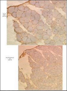
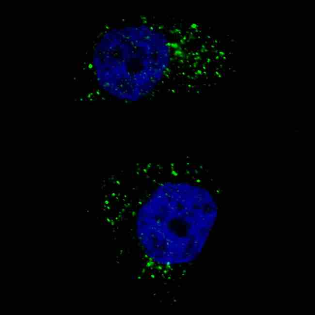
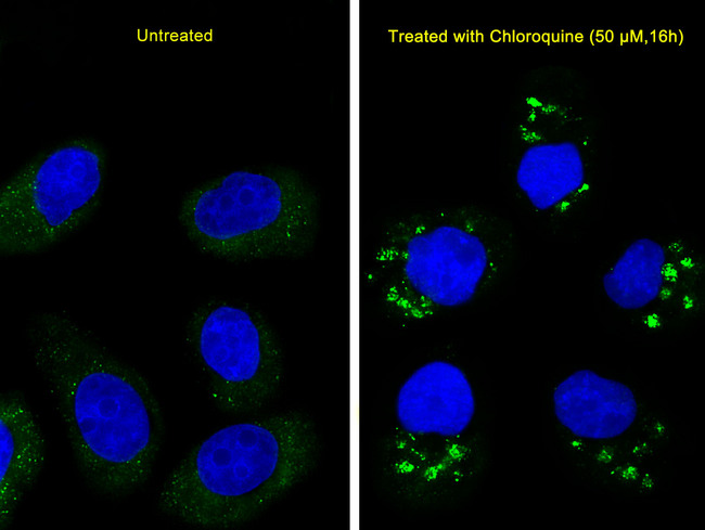
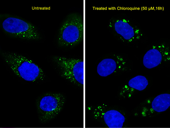
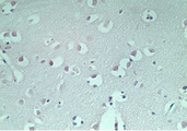
Monoclonal Mouse anti‑Human MAP1LC3A / LC3A Antibody (IHC, IF, WB) LS‑C165694
Monoclonal Mouse anti‑Human MAP1LC3A / LC3A Antibody (IHC, IF, WB) LS‑C165694

Antibody:
MAP1LC3A / LC3A Mouse anti-Human Monoclonal Antibody
Application:
IHC, IF, WB
Reactivity:
Human, Mouse, Rat
Format:
Unconjugated, Unmodified
Toll Free North America
 206-374-1102
For Research Use Only
Overview
Antibody:
MAP1LC3A / LC3A Mouse anti-Human Monoclonal Antibody
Application:
IHC, IF, WB
Reactivity:
Human, Mouse, Rat
Format:
Unconjugated, Unmodified
Specifications
Description
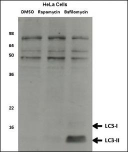
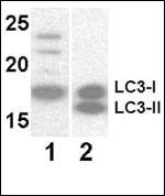
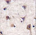
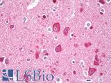
LC3A antibody LS-C165694 is an unconjugated mouse monoclonal antibody to LC3A (MAP1LC3A) from human. It is reactive with human, mouse and rat. Validated for IF, IHC and WB. Cited in 2 publications.
Target
Human MAP1LC3A / LC3A
Synonyms
MAP1LC3A | ATG8E | LC3 | LC3A | MAP1BLC3 | MAP1A/1B light chain 3 A | MAP1A/MAP1B LC3 A | MAP1A/MAP1B light chain 3 A | MAP1ALC3
Host
Mouse
Reactivity
Human, Mouse, Rat
(tested or 100% immunogen sequence identity)
Clonality
IgG1,k
Monoclonal
Conjugations
Unconjugated
Purification
Protein G purified
Modifications
Unmodified
Applications
- IHC (1:50 - 1:100)
- Immunofluorescence (1:200)
- Western blot (1:1000)

|
Performing IHC? See our complete line of Immunohistochemistry Reagents including antigen retrieval solutions, blocking agents
ABC Detection Kits and polymers, biotinylated secondary antibodies, substrates and more.
|
Presentation
PBS, 0.09% Sodium Azide
Storage
Maintain refrigerated at 2°C to 8°C for up to 6 months. For long term storage store at -20°C.
Restrictions
For research use only. Intended for use by laboratory professionals.
About MAP1LC3A / LC3A
LSBio Ratings
MAP1LC3A / LC3A Antibody for IHC, IF/Immunofluorescence, WB/Western LS-C165694 has an LSBio Rating of
Publications (4.1)
Learn more about The LSBio Ratings Algorithm
Publications (2)
Mouse knock-out of IOP1 protein reveals its essential role in mammalian cytosolic iron-sulfur protein biogenesis. Song D, Lee FS. The Journal of biological chemistry. 2011 286:15797-805.
Mitochondria-lysosome membrane contacts are defective in GDAP1-related Charcot-Marie-Tooth disease. Lara Cantarero , Elena Juárez-Escoto , Azahara Civera-Tregón , María Rodríguez-Sanz, Mónica Roldán, Raúl Benítez , Janet Hoenicka , Francesc Palau. The Breast : official journal of the European Society of Mastology. 2021 Jan;29:3589-3605.
Customer Reviews (0)
Featured Products
Species:
Human, Mouse
Applications:
IHC, Western blot, Flow Cytometry, ELISA
Species:
Human, Mouse
Applications:
IHC, Western blot, Flow Cytometry, ELISA
Species:
Human, Mouse, Rat
Applications:
IHC, IHC - Paraffin, Immunofluorescence, Western blot
Species:
Human, Mouse, Rat, Bovine, Opossum, Zebrafish
Applications:
IHC, IHC - Paraffin, Western blot
Species:
Human, Monkey, Mouse, Rat, Bovine, Opossum, Chicken, Xenopus, Pufferfish
Applications:
IHC, IHC - Paraffin, Western blot
Request SDS/MSDS
To request an SDS/MSDS form for this product, please contact our Technical Support department at:
Technical.Support@LSBio.com
Requested From: United States
Date Requested: 4/18/2024
Date Requested: 4/18/2024


