Login
Registration enables users to use special features of this website, such as past
order histories, retained contact details for faster checkout, review submissions, and special promotions.
order histories, retained contact details for faster checkout, review submissions, and special promotions.
Forgot password?
Registration enables users to use special features of this website, such as past
order histories, retained contact details for faster checkout, review submissions, and special promotions.
order histories, retained contact details for faster checkout, review submissions, and special promotions.
Quick Order
Products
Antibodies
ELISA and Assay Kits
Research Areas
Infectious Disease
Resources
Purchasing
Reference Material
Contact Us
Locations
Orders Processing,
Shipping & Receiving,
Warehouse
2 Shaker Rd Suites
B001/B101
Shirley, MA 01464
Production Lab
Floor 6, Suite 620
20700 44th Avenue W
Lynnwood, WA 98036
Telephone Numbers
Tel: +1 (206) 374-1102
Fax: +1 (206) 577-4565
Contact Us
Additional Contact Details
Login
Registration enables users to use special features of this website, such as past
order histories, retained contact details for faster checkout, review submissions, and special promotions.
order histories, retained contact details for faster checkout, review submissions, and special promotions.
Forgot password?
Registration enables users to use special features of this website, such as past
order histories, retained contact details for faster checkout, review submissions, and special promotions.
order histories, retained contact details for faster checkout, review submissions, and special promotions.
Quick Order
| Catalog Number | Size | Price |
|---|---|---|
| LS-B34-50 | 50 µg (1 mg/ml) | $515 |
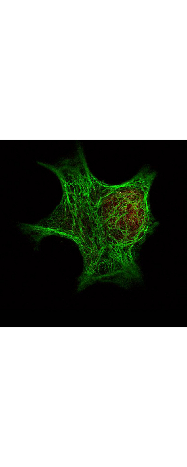
![HDAC1 Antibody - Immunofluorescence Microscopy - HDAC Antibody. Histone deacetylase (HDAC) antibody detects HDAC (colored GREEN) as used in STED immunofluorescence microscopy. Methanol fixed A431 cells were blocked with normal goat serum. The cells were then probed with 0.4 ug/mL final concentration of anti-HDAC and detected with 0.2 ug/mL DyLight488 conjugated Anti-RABBIT IgG [GOAT] secondary antibody. Also shown in this 2-color STED image is an anti-tubulin monoclonal antibody [MOUSE] ( detected with ATTO 425 conjugated anti-MOUSE IgG [GOAT] ( secondary antibody (colored RED).](https://lsbio-7d62.kxcdn.com/image2/ihc-plus-hdac1-antibody-aa450-482-ls-b34/62907_776555.jpg)

![HDAC1 Antibody - Anti-HDAC-1 Antibody - Western Blot. Western blot of Affinity Purified anti-HDAC-1 antibody shows detection of a band at ~65 kD corresponding to human HDAC1 present in a 293 whole cell lysate (arrowhead). Approximately 35 ug of lysate was separated on a 4-20% Tris-HEPES gel by SDS-PAGE and transferred onto nitrocellulose. After blocking the membrane was probed with the primary antibody diluted to 1:1350. Reaction occurred 2 h at room temperature followed by washes and reaction with a 1:10000 dilution of IRDye800 conjugated Rb-a-Goat IgG [H&L] MXHu ( for 45 min at room temperature. IRDye800 fluorescence image was captured using the Odyssey Infrared Imaging System developed by LI-COR. IRDye is a trademark of LI-COR, Inc. Other detection systems will yield similar results.](https://lsbio-7d62.kxcdn.com/image2/ihc-plus-hdac1-antibody-aa450-482-ls-b34/62905_51422.jpg)
1 of 5
2 of 5
3 of 5
4 of 5
5 of 5
IHC‑plus™ Polyclonal Rabbit anti‑Human HDAC1 Antibody (aa450‑482, IHC, IF, WB) LS‑B34
IHC‑plus™ Polyclonal Rabbit anti‑Human HDAC1 Antibody (aa450‑482, IHC, IF, WB) LS‑B34

Note: This antibody replaces LS-C18818, LS-C56878
Antibody:
HDAC1 Rabbit anti-Human Polyclonal (aa450-482) Antibody
Application:
IHC, IHC-P, IF, WB, ELISA
Reactivity:
Human
Format:
Unconjugated, Unmodified
Toll Free North America
 206-374-1102
For Research Use Only
Overview
Antibody:
HDAC1 Rabbit anti-Human Polyclonal (aa450-482) Antibody
Application:
IHC, IHC-P, IF, WB, ELISA
Reactivity:
Human
Format:
Unconjugated, Unmodified
Specifications
Description
HDAC1 antibody LS-B34 is an unconjugated rabbit polyclonal antibody to human HDAC1 (aa450-482). Validated for ELISA, IF, IHC and WB. Tested on 20 paraffin-embedded human tissues.
Target
Human HDAC1
Synonyms
HDAC1 | Histone deacetylase 1 | GON-10 | HD1 | RPD3 | RPD3L1
Host
Rabbit
Reactivity
Human
(tested or 100% immunogen sequence identity)
Predicted
Chimpanzee, Mouse, Rat (at least 90% immunogen sequence identity)
Clonality
IgG
Polyclonal
Conjugations
Unconjugated
Purification
Affinity purified
Modifications
Unmodified
Immunogen
Anti-HDAC-1 antibody was prepared from whole rabbit serum produced by repeated immunizations with a synthetic peptide corresponding to a C-Terminal region near amino acids 450-482 of Human HDAC-1.
Epitope
aa450-482
Specificity
A BLAST analysis was used to suggest reactivity with this protein from human, mouse, rat and chimpanzee sources based on 100% homology for the immunogen sequence. Cross reactivity may occur with HDAC-1 from bovine (82% homology) and chicken (80% homology) sources. Cross reactivity with HDAC-1 homologues from other sources has not been determined.
Applications
- IHC
- IHC - Paraffin (20 µg/ml)
- Immunofluorescence (10 µg/ml)
- Western blot (1:1000 - 1:5000)
- ELISA (1:10000 - 1:50000)

|
Performing IHC? See our complete line of Immunohistochemistry Reagents including antigen retrieval solutions, blocking agents
ABC Detection Kits and polymers, biotinylated secondary antibodies, substrates and more.
|
Usage
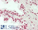
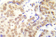
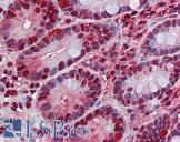
Immunohistochemistry: LS-B34 was validated for use in immunohistochemistry on a panel of 21 formalin-fixed, paraffin-embedded (FFPE) human tissues after heat induced antigen retrieval in pH 6.0 citrate buffer. After incubation with the primary antibody, slides were incubated with biotinylated secondary antibody, followed by alkaline phosphatase-streptavidin and chromogen. The stained slides were evaluated by a pathologist to confirm staining specificity. The optimal working concentration for LS-B34 was determined to be 20 ug/ml.
Presentation
0.02 M Potassium Phosphate, pH 7.2, 0.15 M NaCl, 0.01% Sodium Azide
Storage
Store at 4°C or -20°C. Avoid freeze-thaw cycles.
Restrictions
For research use only. Intended for use by laboratory professionals.
About HDAC1
Validation
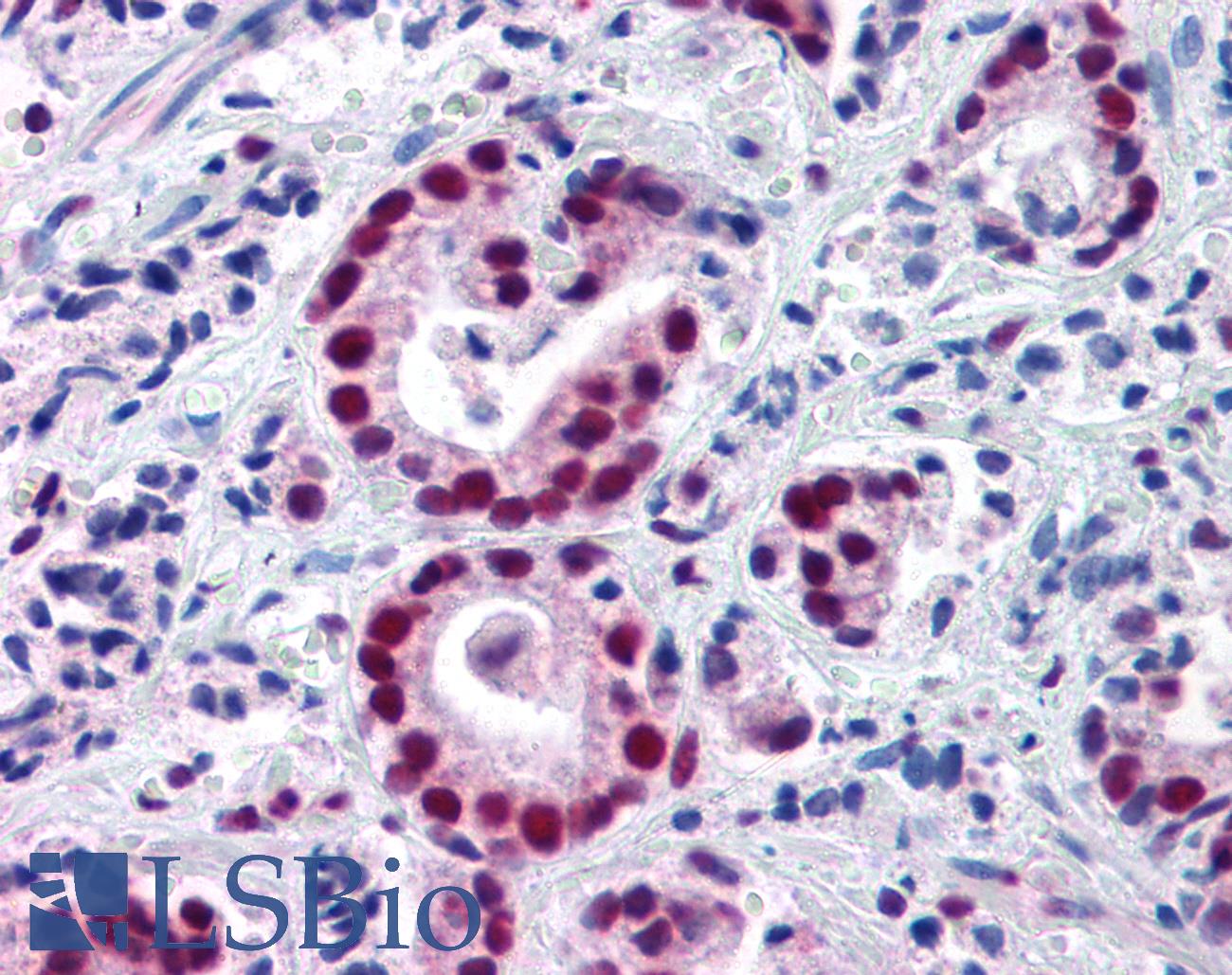
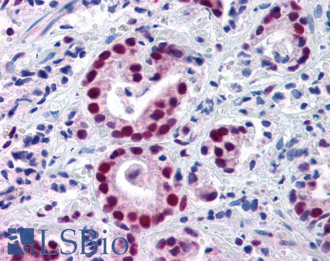
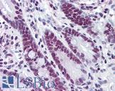
Anti-HDAC1 antibody IHC of human prostate carcinoma. Immunohistochemistry of formalin-fixed, paraffin-embedded tissue after heat-induced antigen retrieval. Antibody concentration 20 ug/ml.
Anti-HDAC1 antibody IHC of human prostate carcinoma. Immunohistochemistry of formalin-fixed, paraffin-embedded tissue after heat-induced antigen retrieval. Antibody concentration 20 ug/ml.
See More About...
LSBio Ratings
IHC-plus™ HDAC1 Antibody (aa450-482) for IHC, IF/Immunofluorescence, WB/Western, ELISA LS-B34 has an LSBio Rating of
Laboratory Validation Score (4)
Learn more about The LSBio Ratings Algorithm
Publications (0)
Customer Reviews (0)
Featured Products
Species:
Human, Mouse, Rat
Applications:
IHC, IHC - Paraffin, Immunofluorescence, Western blot, ELISA
Species:
Human, Monkey, Mouse, Rat, Bovine, Dog, Guinea pig, Hamster, Horse, Rabbit
Applications:
Western blot, Chromatin Immunoprecipitation, Peptide Enzyme-Linked Immunosorbent Assay
Species:
Human, Mouse, Rat
Applications:
IHC, IHC - Paraffin, Western blot
Species:
Human, Mouse, Rat
Applications:
IHC, IHC - Paraffin
Species:
Human
Applications:
IHC, IHC - Paraffin, Immunofluorescence, Western blot, ELISA
Request SDS/MSDS
To request an SDS/MSDS form for this product, please contact our Technical Support department at:
Technical.Support@LSBio.com
Requested From: United States
Date Requested: 4/19/2024
Date Requested: 4/19/2024


