Login
Registration enables users to use special features of this website, such as past
order histories, retained contact details for faster checkout, review submissions, and special promotions.
order histories, retained contact details for faster checkout, review submissions, and special promotions.
Forgot password?
Registration enables users to use special features of this website, such as past
order histories, retained contact details for faster checkout, review submissions, and special promotions.
order histories, retained contact details for faster checkout, review submissions, and special promotions.
Quick Order
Products
Antibodies
ELISA and Assay Kits
Research Areas
Infectious Disease
Resources
Purchasing
Reference Material
Contact Us
Locations
Orders Processing,
Shipping & Receiving,
Warehouse
2 Shaker Rd Suites
B001/B101
Shirley, MA 01464
Production Lab
Floor 6, Suite 620
20700 44th Avenue W
Lynnwood, WA 98036
Telephone Numbers
Tel: +1 (206) 374-1102
Fax: +1 (206) 577-4565
Contact Us
Additional Contact Details
Login
Registration enables users to use special features of this website, such as past
order histories, retained contact details for faster checkout, review submissions, and special promotions.
order histories, retained contact details for faster checkout, review submissions, and special promotions.
Forgot password?
Registration enables users to use special features of this website, such as past
order histories, retained contact details for faster checkout, review submissions, and special promotions.
order histories, retained contact details for faster checkout, review submissions, and special promotions.
Quick Order
| Catalog Number | Size | Price |
|---|---|---|
| LS-C166517-200 | 200 µl (0.5 mg/ml) | $393 |
1 of 12
2 of 12
3 of 12
4 of 12
5 of 12
6 of 12
7 of 12
8 of 12
9 of 12
10 of 12
11 of 12
12 of 12
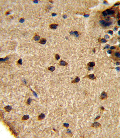
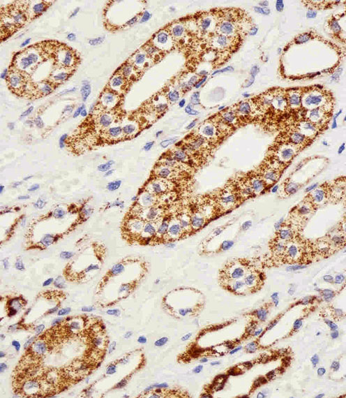
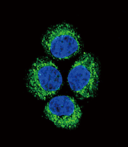
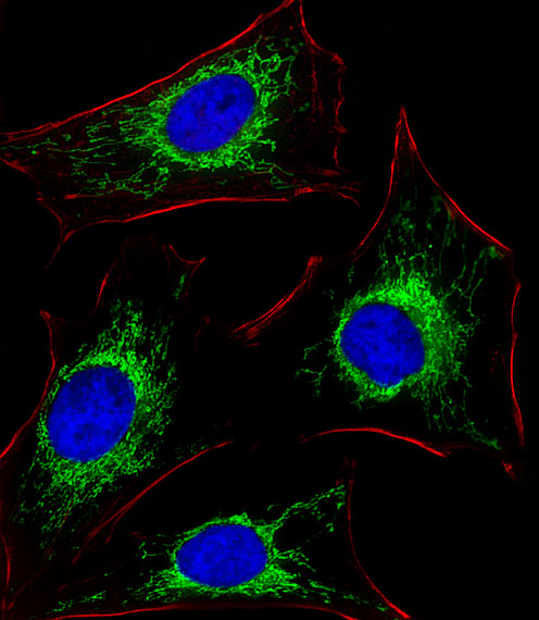
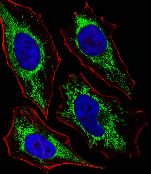
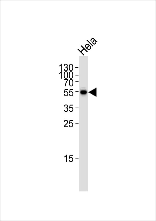
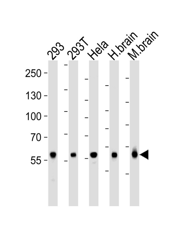
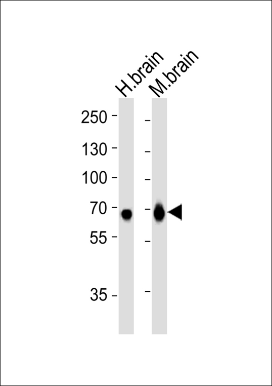
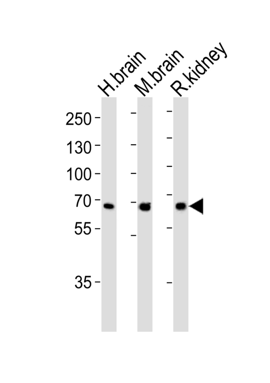
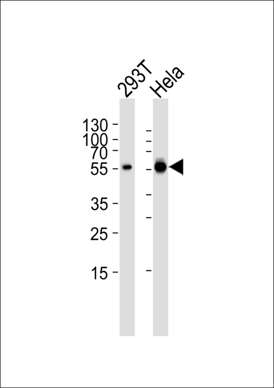
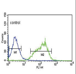
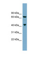
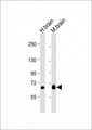
Polyclonal Rabbit anti‑Human GLS / Glutaminase Antibody (aa516‑545, IHC, IF, WB) LS‑C166517
Polyclonal Rabbit anti‑Human GLS / Glutaminase Antibody (aa516‑545, IHC, IF, WB) LS‑C166517
Antibody:
GLS / Glutaminase Rabbit anti-Human Polyclonal (aa516-545) Antibody
Application:
IHC, IHC-P, IF, WB, Flo
Reactivity:
Human, Mouse, Rat
Format:
Unconjugated, Unmodified
Toll Free North America
 206-374-1102
For Research Use Only
Overview
Antibody:
GLS / Glutaminase Rabbit anti-Human Polyclonal (aa516-545) Antibody
Application:
IHC, IHC-P, IF, WB, Flo
Reactivity:
Human, Mouse, Rat
Format:
Unconjugated, Unmodified
Specifications
Description
Glutaminase antibody LS-C166517 is an unconjugated rabbit polyclonal antibody to Glutaminase (GLS) (aa516-545) from human. It is reactive with human, mouse and rat. Validated for Flow, IF, IHC and WB.
Target
Human GLS / Glutaminase
Synonyms
GLS | AAD20 | GLS1 | GAM | Glutaminase C | GAC | K-glutaminase | KIAA0838 | L-glutamine amidohydrolase | Glutaminase | KGA
Host
Rabbit
Reactivity
Human, Mouse, Rat
(tested or 100% immunogen sequence identity)
Predicted
Rat (at least 90% immunogen sequence identity)
Clonality
Polyclonal
Conjugations
Unconjugated
Purification
Protein A purified
Modifications
Unmodified
Epitope
aa516-545
Specificity
This GLS antibody is generated from rabbits immunized with a KLH conjugated synthetic peptide between 516-545 amino acids from the C-terminal region of human GLS.
Applications
- IHC
- IHC - Paraffin (1:10 - 1:50)
- Immunofluorescence (1:10 - 1:50)
- Western blot (1:1000)
- Flow Cytometry (1:10 - 1:50)

|
Performing IHC? See our complete line of Immunohistochemistry Reagents including antigen retrieval solutions, blocking agents
ABC Detection Kits and polymers, biotinylated secondary antibodies, substrates and more.
|
Presentation
PBS, 0.09% Sodium Azide
Storage
Maintain refrigerated at 2°C to 8°C for up to 6 months. For long term storage store at -20°C.
Restrictions
For research use only. Intended for use by laboratory professionals.
About GLS / Glutaminase
Publications (0)
Customer Reviews (0)
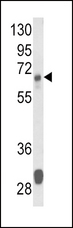
Featured Products
Species:
Mouse, Human
Applications:
Western blot
![GLS / Glutaminase Antibody - Immunofluorescence of monoclonal antibody to GLS on HeLa cell. [antibody concentration 10 ug/ml]](https://lsbio-7d62.kxcdn.com//image2/gls-glutaminase-antibody-clone-5c4-ls-c197311/145039_727617.jpg)
Species:
Human
Applications:
Immunofluorescence, Western blot, ELISA
Species:
Human, Mouse
Applications:
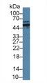
IHC, Immunofluorescence, Western blot, Flow Cytometry, ELISA
Species:
Human, Mouse
Applications:
Western blot, Peptide Enzyme-Linked Immunosorbent Assay
Reactivity:
Mouse
Range:
0.313-20 ng/ml
Request SDS/MSDS
To request an SDS/MSDS form for this product, please contact our Technical Support department at:
Technical.Support@LSBio.com
Requested From: United States
Date Requested: 4/23/2024
Date Requested: 4/23/2024


