order histories, retained contact details for faster checkout, review submissions, and special promotions.
Forgot password?
order histories, retained contact details for faster checkout, review submissions, and special promotions.
Locations
Orders Processing,
Shipping & Receiving,
Warehouse
2 Shaker Rd Suites
B001/B101
Shirley, MA 01464
Production Lab
Floor 6, Suite 620
20700 44th Avenue W
Lynnwood, WA 98036
Telephone Numbers
Tel: +1 (206) 374-1102
Fax: +1 (206) 577-4565
Contact Us
Additional Contact Details
order histories, retained contact details for faster checkout, review submissions, and special promotions.
Forgot password?
order histories, retained contact details for faster checkout, review submissions, and special promotions.
GFP
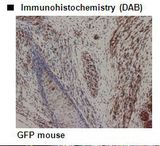
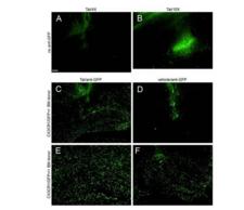
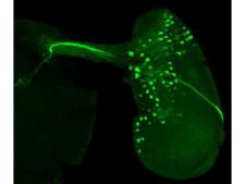
Green Fluorescent Protein (GFP)
Green Fluorescent Protein (GFP) exhibits bright green fluorescence when exposed to blue light. Although many other marine organisms have similar green fluorescent proteins, GFP traditionally refers to the protein first isolated from the jellyfish Aequorea victoria. GFP from A. victoria has a major excitation peak at a wavelength of 395 nm and a minor one at 475 nm. Its emission peak is at 509 nm, which is in the lower green portion of the visible spectrum. GFP from the sea pansy (Renilla reniformis) has a single major excitation peak at 498 nm. In cell and molecular biology, the GFP gene is frequently used as a reporter of expression. In modified forms it has been used to make biosensors, and many animals have been created that express GFP as a proof-of-concept that a gene can be expressed throughout a given organism. The GFP gene can be introduced into organisms and maintained in their genome through breeding, injection with a viral vector, or cell transformation.
Publications (32)
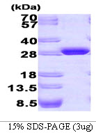
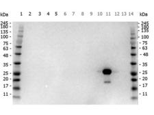
![GFP Antibody - Anti-GFP Antibody - Western Blot. Western blot of GFP recombinant protein detected with monoclonal anti-GFP antibody. GFP recombinant protein was expressed in HeLa cells, where 50 ng (lane 1), 100 ng (lane 2) and 500 ng (lane 3) of lysate were loaded per lane. Mab anti-GFP detects a 27 kD band corresponding to the epitope tag GFP. The cell lysates were prepared in a RIPA buffer containing 200 mM NaCl. A 4-12% Bis-Tris gradient gel (Invitrogen) was used for SDS-PAGE. The protein was transferred to nitrocellulose using standard methods. After blocking with 5% BLOTTO in PBS, the membrane was probed with the primary antibody diluted to 1.0 mg/ml for 1 h at room temperature followed by washes and reaction with a 1:2500 dilution of IRDye 800 conjugated Goat-a-Mouse IgG [H&L] MX10 (. IRDye 800 fluorescence image was captured using the Odyssey Infrared Imaging System developed by LI-COR. IRDye is a trademark of LI-COR, Inc. Other detection systems will yield similar results.](https://lsbio-7d62.kxcdn.com/image2/gfp-antibody-clone-9f9.f9-ls-c154208/81891_5149892.jpg)

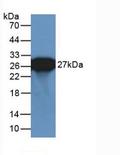
![GFP Antibody - Anti-GFP Antibody - Western Blot. Western blot of GFP protein detected with polyclonal anti-GFP antibody. Wild type GFP (0.1 ug) was used to spike 30 ug of a HeLa whole cell lysate. This antibody detects a 27 kD band corresponding to the epitope tag GFP. A 4-20% Tris-Glycine gradient gel was used for SDS-PAGE. The protein was transferred to nitrocellulose using standard methods. After blocking with 5% BLOTTO in PBS, the membrane was probed overnight at 4C with the primary antibody diluted in 5% BLOTTO to 1:1000, followed by washes and reaction with a 1:10000 dilution of IRDye 800 conjugated Goat-a-Rabbit IgG [H&L] MX10 (. IRDye 800 fluorescence image was captured using the Odyssey Infrared Imaging System developed by LI-COR. IRDye is a trademark of LI-COR, Inc. Other detection systems will yield similar results.](https://lsbio-7d62.kxcdn.com/image2/gfp-antibody-ls-c154219/81899_5149904.jpg)


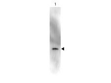
![GFP Antibody - Anti-GFP Antibody - Western Blot. Western blot of GFP recombinant protein detected with polyclonal anti-GFP antibody. Lane 1 shows detection of a 33 kD band corresponding to a GFP containing recombinant protein (arrowhead) expressed in HeLa cells. Lane 2 shows no staining of a mock transfected HeLa cell lysate. A 4-12% Bis-Tris gradient gel was used for SDS-PAGE. The protein was transferred to nitrocellulose using standard methods. After blocking the membrane was probed with the primary antibody diluted to 1 ug/ml for 1 h at room temperature followed by washes and reaction with a 1:2500 dilution of IRDye 800 conjugated Donkey-a-Goat IgG [H&L] MX7 (. The IRDye 800 fluorescence image was captured using the Odyssey Infrared Imaging System developed by LI-COR. IRDye is a trademark of LI-COR, Inc. Other detection systems will yield similar results. This image was taken for the unconjugated form of this product. Other forms have not been tested.](https://lsbio-7d62.kxcdn.com/image2/gfp-antibody-fitc-ls-c154186/81887_5149889.jpg)
If you do not find the reagent or information you require, please contact Customer.Support@LSBio.com to inquire about additional products in development.


