Login
Registration enables users to use special features of this website, such as past
order histories, retained contact details for faster checkout, review submissions, and special promotions.
order histories, retained contact details for faster checkout, review submissions, and special promotions.
Forgot password?
Registration enables users to use special features of this website, such as past
order histories, retained contact details for faster checkout, review submissions, and special promotions.
order histories, retained contact details for faster checkout, review submissions, and special promotions.
Quick Order
Products
Antibodies
ELISA and Assay Kits
Research Areas
Infectious Disease
Resources
Purchasing
Reference Material
Contact Us
Locations
Orders Processing,
Shipping & Receiving,
Warehouse
2 Shaker Rd Suites
B001/B101
Shirley, MA 01464
Production Lab
Floor 6, Suite 620
20700 44th Avenue W
Lynnwood, WA 98036
Telephone Numbers
Tel: +1 (206) 374-1102
Fax: +1 (206) 577-4565
Contact Us
Additional Contact Details
Login
Registration enables users to use special features of this website, such as past
order histories, retained contact details for faster checkout, review submissions, and special promotions.
order histories, retained contact details for faster checkout, review submissions, and special promotions.
Forgot password?
Registration enables users to use special features of this website, such as past
order histories, retained contact details for faster checkout, review submissions, and special promotions.
order histories, retained contact details for faster checkout, review submissions, and special promotions.
Quick Order
| Catalog Number | Size | Price |
|---|---|---|
| LS-C46139-0.1 | 0.1 mg (1 mg/ml) | $316 |
1 of 2
2 of 2
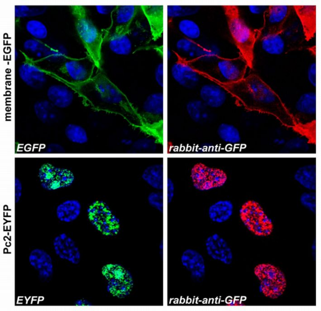
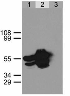
Polyclonal Rabbit anti‑Aequorea victoria GFP Antibody (clone Polyclonal, WB) LS‑C46139
Polyclonal Rabbit anti‑Aequorea victoria GFP Antibody (clone Polyclonal, WB) LS‑C46139
Antibody:
GFP Rabbit anti-Aequorea victoria Polyclonal (Polyclonal) Antibody
Application:
ICC, WB, IP
Reactivity:
Aequorea victoria, All species
Format:
Unconjugated, Unmodified
Toll Free North America
 206-374-1102
For Research Use Only
Overview
Antibody:
GFP Rabbit anti-Aequorea victoria Polyclonal (Polyclonal) Antibody
Application:
ICC, WB, IP
Reactivity:
Aequorea victoria, All species
Format:
Unconjugated, Unmodified
Specifications
Description
GFP antibody LS-C46139 is an unconjugated rabbit polyclonal antibody to all species GFP. Validated for ICC, IP and WB.
Host
Rabbit
Reactivity
Aequorea victoria, All species
Clonality
Polyclonal
Clone
Polyclonal
Conjugations
Unconjugated
Purification
Purified from rabbit serum by affinity chromatography. Greater than 95% by SDS-PAGE.
Modifications
Unmodified
Immunogen
EGFP, a native full-length protein.
Specificity
The polyclonal antibody recognizes GFP, EGFP, EYFP fusion proteins in all species.
Applications
- ICC
- Western blot (0.5 - 1.5 µg/ml)
- Immunoprecipitation
Presentation
PBS, pH 7.4, 15 mM Sodium Azide
Storage
Store at 2°C to 8°C. Do not freeze.
Restrictions
For research use only. Intended for use by laboratory professionals.
Publications (0)
Customer Reviews (0)
Featured Products
Species:
Aequorea victoria
Applications:
IHC - Paraffin, IHC - Frozen, Western blot, ELISA
Species:
Aequorea victoria
Applications:
IHC, IHC - Paraffin, Western blot, Immunoprecipitation, ELISA
Species:
Aequorea victoria
Applications:
IHC, IHC - Paraffin, IHC - Frozen, Western blot, ELISA
Species:
Aequorea victoria
Applications:
IHC, Immunofluorescence, Western blot, ELISA

Species:
Aequorea victoria
Applications:
IHC, Immunofluorescence, Western blot, Flow Cytometry, ELISA
Request SDS/MSDS
To request an SDS/MSDS form for this product, please contact our Technical Support department at:
Technical.Support@LSBio.com
Requested From: United States
Date Requested: 4/19/2024
Date Requested: 4/19/2024


