order histories, retained contact details for faster checkout, review submissions, and special promotions.
Forgot password?
order histories, retained contact details for faster checkout, review submissions, and special promotions.
Locations
Orders Processing,
Shipping & Receiving,
Warehouse
2 Shaker Rd Suites
B001/B101
Shirley, MA 01464
Production Lab
Floor 6, Suite 620
20700 44th Avenue W
Lynnwood, WA 98036
Telephone Numbers
Tel: +1 (206) 374-1102
Fax: +1 (206) 577-4565
Contact Us
Additional Contact Details
order histories, retained contact details for faster checkout, review submissions, and special promotions.
Forgot password?
order histories, retained contact details for faster checkout, review submissions, and special promotions.
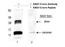
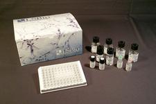
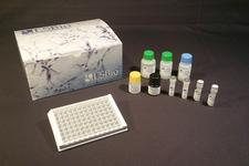
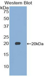
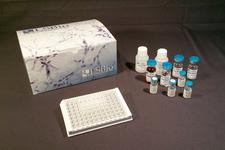
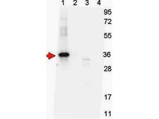
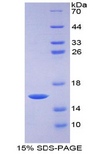
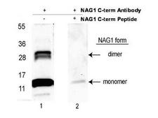
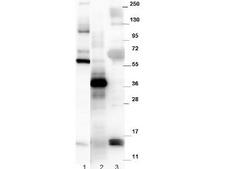
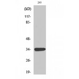
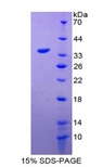
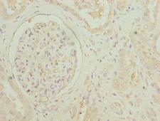
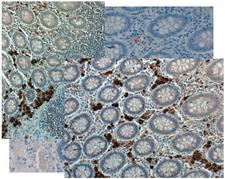
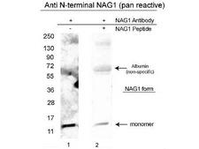
GDF15
growth differentiation factor 15
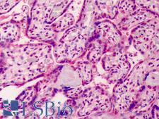
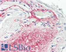
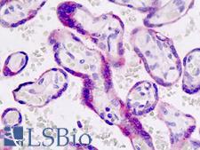
Growth differentiation factor 15 (GDF15) is a protein belonging to the transforming growth factor beta superfamily that has a role in regulating inflammatory and apoptotic pathways in injured tissues and during disease processes. GDF15 is also known as TGF-PL, MIC-1, PDF, PLAB, and PTGFB. GDF15 mRNA is most abundant in the liver, with lower levels seen in some other tissues. Its expression in liver can be significantly up-regulated in during injury of organs such as liver, kidney, heart and lung.
| Gene Name: | growth differentiation factor 15 |
| Family/Subfamily: | TGF beta , not assigned-TGF beta |
| Synonyms: | GDF15, MIC1, MIC-1, NAG-1, NRG-1, NSAID-regulated gene 1 protein, PLAB, NSAID-activated gene 1 protein, Placental TGF-beta, PTGFB, GDF-15, PTGF-beta |
| Target Sequences: | NM_004864 NP_004855.2 Q99988 |
Publications (1)


If you do not find the reagent or information you require, please contact Customer.Support@LSBio.com to inquire about additional products in development.


