Login
Registration enables users to use special features of this website, such as past
order histories, retained contact details for faster checkout, review submissions, and special promotions.
order histories, retained contact details for faster checkout, review submissions, and special promotions.
Forgot password?
Registration enables users to use special features of this website, such as past
order histories, retained contact details for faster checkout, review submissions, and special promotions.
order histories, retained contact details for faster checkout, review submissions, and special promotions.
Quick Order
Products
Antibodies
ELISA and Assay Kits
Research Areas
Infectious Disease
Resources
Purchasing
Reference Material
Contact Us
Locations
Orders Processing,
Shipping & Receiving,
Warehouse
2 Shaker Rd Suites
B001/B101
Shirley, MA 01464
Production Lab
Floor 6, Suite 620
20700 44th Avenue W
Lynnwood, WA 98036
Telephone Numbers
Tel: +1 (206) 374-1102
Fax: +1 (206) 577-4565
Contact Us
Additional Contact Details
Login
Registration enables users to use special features of this website, such as past
order histories, retained contact details for faster checkout, review submissions, and special promotions.
order histories, retained contact details for faster checkout, review submissions, and special promotions.
Forgot password?
Registration enables users to use special features of this website, such as past
order histories, retained contact details for faster checkout, review submissions, and special promotions.
order histories, retained contact details for faster checkout, review submissions, and special promotions.
Quick Order
| Catalog Number | Size | Price |
|---|---|---|
| LS-B2996-50 | 50 µg (0.5 mg/ml) | $485 |
1 of 7
2 of 7
3 of 7
4 of 7
5 of 7
6 of 7
7 of 7
PathPlus™ Polyclonal Goat anti‑Human FOXC1 Antibody (aa541‑553, IHC, IF, WB) LS‑B2996
PathPlus™ Polyclonal Goat anti‑Human FOXC1 Antibody (aa541‑553, IHC, IF, WB) LS‑B2996

Note: This antibody replaces LS-C54851, LS-C66329
Antibody:
FOXC1 Goat anti-Human Polyclonal (aa541-553) Antibody
Application:
IHC, IHC-P, IF, WB, Peptide-ELISA
Reactivity:
Human, Monkey, Mouse, Xenopus, Zebrafish
Format:
Unconjugated, Unmodified
Toll Free North America
 206-374-1102
For Research Use Only
Overview
Antibody:
FOXC1 Goat anti-Human Polyclonal (aa541-553) Antibody
Application:
IHC, IHC-P, IF, WB, Peptide-ELISA
Reactivity:
Human, Monkey, Mouse, Xenopus, Zebrafish
Format:
Unconjugated, Unmodified
Specifications
Description
FOXC1 is a forkhead transcription factor involved in embryogenesis, cardiac morphogenesis and ocular development. Inherited mutations in FOXC1 lead to glaucoma, Axenfeld-Rieger syndrome and cerebral small-vessel disease (CSVD), described by increased risk for stroke and progressive cognitive decline. Upregulation of FOXC1 in cancer is common and linked with poor prognosis, as it leads to increased expression of vimentin, N-cadherin and fibronectin and consequently promotes migration and metastasis. In immunohistochemistry, FOXC1 has highest cytoplasmic and nuclear positivity in the central nervous system and salivary gland, and is also moderately expressed in most other tissues throughout the body.
References: Developmental biology. 2006. 421-436. ISBN-10: 0-87893-243-7; Molecular Medicine Reports. 12 (6): 8003–9, PMID: 26461269; Oncogene. 2017 Jul 13;36(28):3957-3963, PMID: 28288141; J Clin Invest. 2014 Nov;124(11):4877-81, PMID: 25250569
Target
Human FOXC1
Synonyms
FOXC1 | Forkhead-like 7 | FREAC3 | FKHL7 | Forkhead-related activator 3 | Forkhead-related protein FKHL7 | IGDA | IHG1 | IRID1 | Forkhead box protein C1 | Mesenchyme fork head protein 1 | Myeloid factor-delta | ARA | Forkhead box C1 | FREAC-3 | RIEG3
Host
Goat
Reactivity
Human, Monkey, Mouse, Xenopus, Zebrafish
(tested or 100% immunogen sequence identity)
Clonality
Polyclonal
Conjugations
Unconjugated
Purification
Purified from goat serum by ammonium sulphate precipitation followed by antigen affinity chromatography using the immunizing peptide.
Modifications
Unmodified
Immunogen
Peptide with sequence RTSGAFVYDCSKF, from the C-Terminus of protein sequence according to NP_001444.2.
Epitope
aa541-553
Specificity
Human FOXC1.
Applications
- IHC
- IHC - Paraffin (3.75 µg/ml)
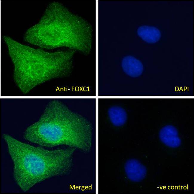
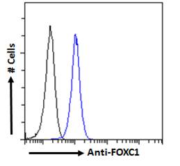
- Immunofluorescence (10 µg/ml)
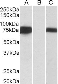
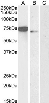
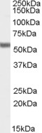
- Western blot (0.5 - 1 µg/ml)
- Peptide Enzyme-Linked Immunosorbent Assay (1:32000)

|
Performing IHC? See our complete line of Immunohistochemistry Reagents including antigen retrieval solutions, blocking agents
ABC Detection Kits and polymers, biotinylated secondary antibodies, substrates and more.
|
Usage
Immunohistochemistry: LS-B2996 was validated for use in immunohistochemistry on a panel of 21 formalin-fixed, paraffin-embedded (FFPE) human tissues after heat induced antigen retrieval in pH 6.0 citrate buffer. After incubation with the primary antibody, slides were incubated with biotinylated secondary antibody, followed by alkaline phosphatase-streptavidin and chromogen. The stained slides were evaluated by a pathologist to confirm staining specificity. The optimal working concentration for LS-B2996 was determined to be 3.75 ug/ml.
Presentation
TBS, pH 7.3, 0.02% Sodium Azide, 0.5% BSA
Storage
Aliquot and store at -20°C. Avoid freeze-thaw cycles.
Restrictions
For research use only. Intended for use by laboratory professionals.
About FOXC1
Validation
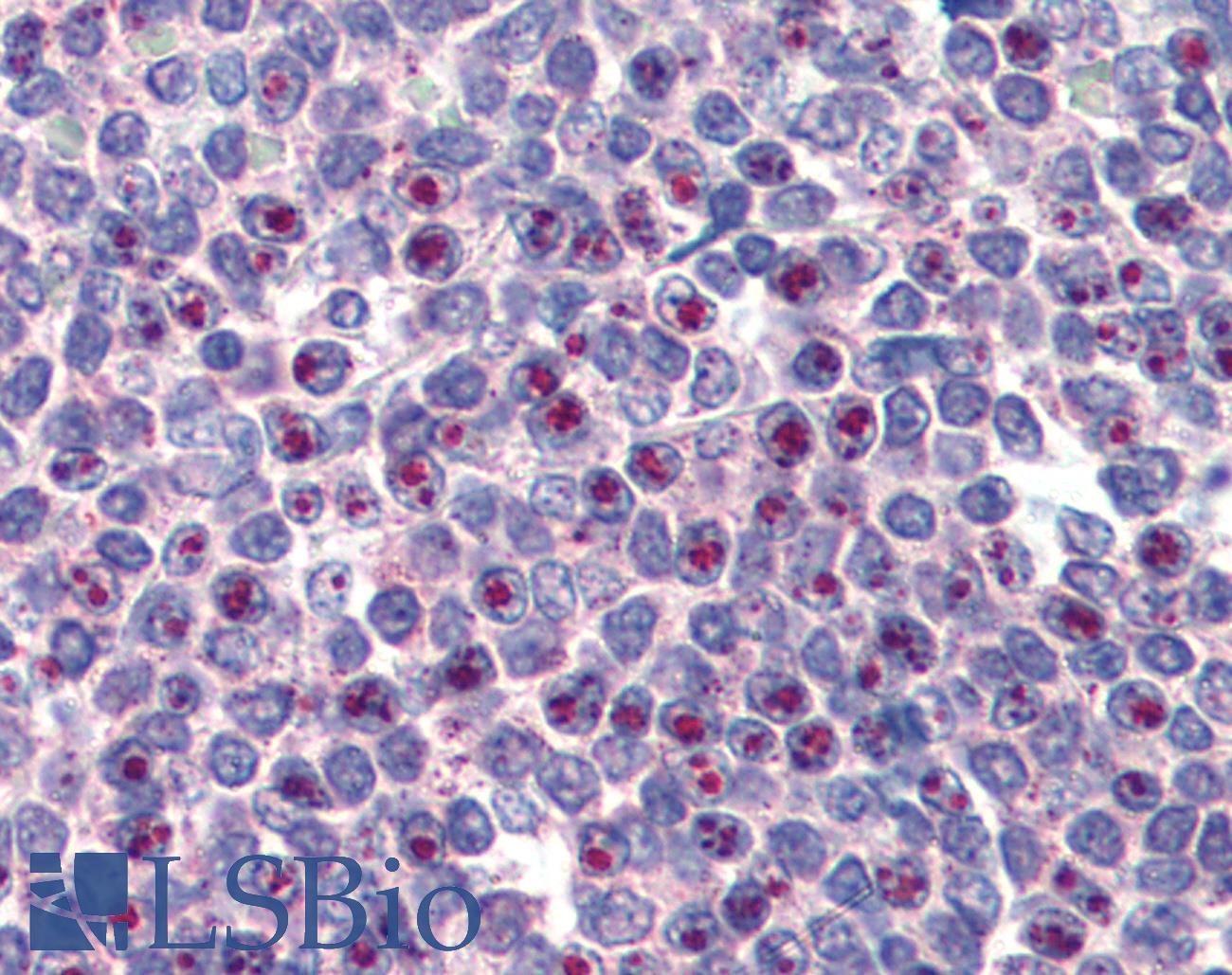
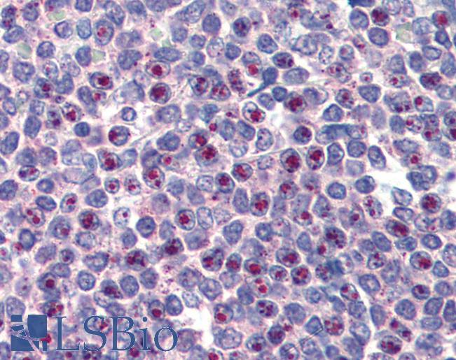
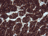
Anti-FOXC1 antibody IHC of human spleen. Immunohistochemistry of formalin-fixed, paraffin-embedded tissue after heat-induced antigen retrieval. Antibody concentration 75 ug/ml.
Anti-FOXC1 antibody IHC of human spleen. Immunohistochemistry of formalin-fixed, paraffin-embedded tissue after heat-induced antigen retrieval. Antibody concentration 75 ug/ml.
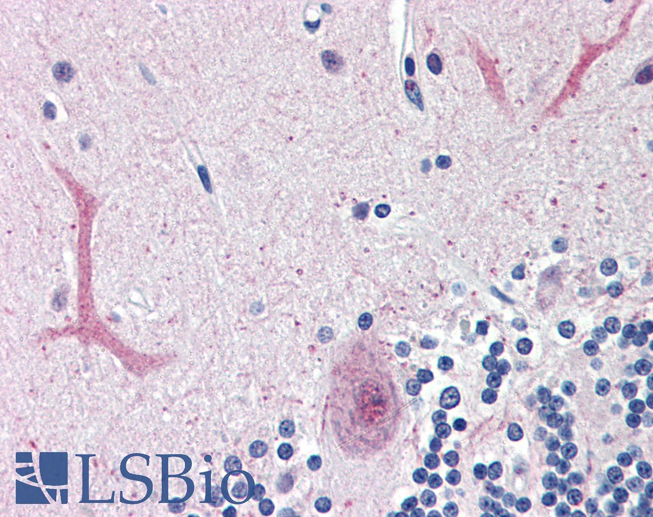
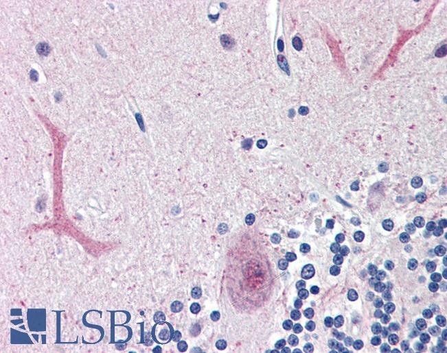
Anti-FOXC1 antibody IHC of human brain, cerebellum. Immunohistochemistry of formalin-fixed, paraffin-embedded tissue after heat-induced antigen retrieval. Antibody concentration 75 ug/ml.
Anti-FOXC1 antibody IHC of human brain, cerebellum. Immunohistochemistry of formalin-fixed, paraffin-embedded tissue after heat-induced antigen retrieval. Antibody concentration 75 ug/ml.
See More About...
LSBio Ratings
PathPlus™ FOXC1 Antibody (aa541-553) for IHC, IF/Immunofluorescence, WB/Western LS-B2996 has an LSBio Rating of
Laboratory Validation Score (5)
Learn more about The LSBio Ratings Algorithm
Publications (0)
Customer Reviews (0)
Featured Products
Species:
Human
Applications:
IHC, Immunofluorescence, Western blot, Flow Cytometry

Reactivity:
Mouse
Range:
78-5000 pg/ml
Reactivity:
Human
Range:
6.25-400 pg/ml

Reactivity:
All species
Range:
123.5-10000 pg/ml
Request SDS/MSDS
To request an SDS/MSDS form for this product, please contact our Technical Support department at:
Technical.Support@LSBio.com
Requested From: United States
Date Requested: 4/18/2024
Date Requested: 4/18/2024


