Login
Registration enables users to use special features of this website, such as past
order histories, retained contact details for faster checkout, review submissions, and special promotions.
order histories, retained contact details for faster checkout, review submissions, and special promotions.
Forgot password?
Registration enables users to use special features of this website, such as past
order histories, retained contact details for faster checkout, review submissions, and special promotions.
order histories, retained contact details for faster checkout, review submissions, and special promotions.
Quick Order
Products
Antibodies
ELISA and Assay Kits
Research Areas
Infectious Disease
Resources
Purchasing
Reference Material
Contact Us
Locations
Orders Processing,
Shipping & Receiving,
Warehouse
2 Shaker Rd Suites
B001/B101
Shirley, MA 01464
Production Lab
Floor 6, Suite 620
20700 44th Avenue W
Lynnwood, WA 98036
Telephone Numbers
Tel: +1 (206) 374-1102
Fax: +1 (206) 577-4565
Contact Us
Additional Contact Details
Login
Registration enables users to use special features of this website, such as past
order histories, retained contact details for faster checkout, review submissions, and special promotions.
order histories, retained contact details for faster checkout, review submissions, and special promotions.
Forgot password?
Registration enables users to use special features of this website, such as past
order histories, retained contact details for faster checkout, review submissions, and special promotions.
order histories, retained contact details for faster checkout, review submissions, and special promotions.
Quick Order
| Catalog Number | Size | Price |
|---|---|---|
| LS-B6446-50 | 50 µg (0.5 mg/ml) | $485 |

1 of 10
2 of 10
3 of 10
4 of 10
5 of 10
6 of 10
7 of 10
8 of 10
9 of 10
10 of 10
IHC‑plus™ Polyclonal Goat anti‑Human DAP3 Antibody (aa387‑398, IHC, IF, WB) LS‑B6446
IHC‑plus™ Polyclonal Goat anti‑Human DAP3 Antibody (aa387‑398, IHC, IF, WB) LS‑B6446

Note: This antibody replaces LS-C34641
Antibody:
DAP3 Goat anti-Human Polyclonal (aa387-398) Antibody
Application:
IHC, IHC-P, IF, WB, Peptide-ELISA
Reactivity:
Human, Monkey
Format:
Unconjugated, Unmodified
Toll Free North America
 206-374-1102
For Research Use Only
Overview
Antibody:
DAP3 Goat anti-Human Polyclonal (aa387-398) Antibody
Application:
IHC, IHC-P, IF, WB, Peptide-ELISA
Reactivity:
Human, Monkey
Format:
Unconjugated, Unmodified
Specifications
Description
DAP3 antibody LS-B6446 is an unconjugated goat polyclonal antibody to DAP3 (aa387-398) from human. It is reactive with human and monkey. Validated for IF, IHC, Peptide-ELISA and WB. Tested on 20 paraffin-embedded human tissues.
Target
Human DAP3
Synonyms
DAP3 | Death associated protein 3 | Death-associated protein 3 | MRP-S29 | BMRP-10 | DAP-3 | MRPS29 | S29mt
Host
Goat
Reactivity
Human, Monkey
(tested or 100% immunogen sequence identity)
Clonality
Polyclonal
Conjugations
Unconjugated
Purification
Purified from goat serum by ammonium sulphate precipitation followed by antigen affinity chromatography using the immunizing peptide.
Modifications
Unmodified
Immunogen
Peptide with sequence NPSLLERHCAYL, from the C-Terminus of protein sequence according to NP_387506.1NP_001186779.1NP_001186780.1.
Epitope
aa387-398
Specificity
Human DAP3. This antibody is expected to recognise isoform 1 (NP_387506.1), isoform 2 (NP_001186779.1) and isoform 3 (NP_001186780.1). Reported variants represent identical protein (NP_387506.1; NP_004623.1; NP_001186778.1).
Applications
- IHC
- IHC - Paraffin (2.5 - 3.75 µg/ml)
- Immunofluorescence (10 µg/ml)
- Western blot (0.1 - 1 µg/ml)
- Peptide Enzyme-Linked Immunosorbent Assay (1:64000)

|
Performing IHC? See our complete line of Immunohistochemistry Reagents including antigen retrieval solutions, blocking agents
ABC Detection Kits and polymers, biotinylated secondary antibodies, substrates and more.
|
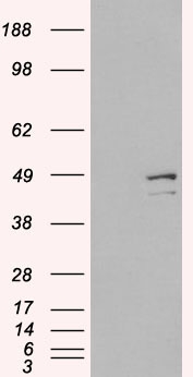
Usage
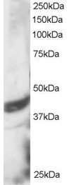
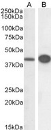
Peptide ELISA: antibody detection limit dilution 1:16000. Western blot: Approx 40 kDa band observed in lysates of cell line HeLa (calculated MW of 45.6 kDa according to NP_387506.1). In transfected HEK293 transiently expressing DAP3 a band of approx. 48 kDa is observed. This band is not observed in the non-transfected HEK293. Recommended concentration: 1-3 ug/ml.
Presentation
TBS, pH 7.3, 0.02% Sodium Azide, 0.5% BSA
Storage
Aliquot and store at -20°C. Avoid freeze-thaw cycles.
Restrictions
For research use only. Intended for use by laboratory professionals.
About DAP3
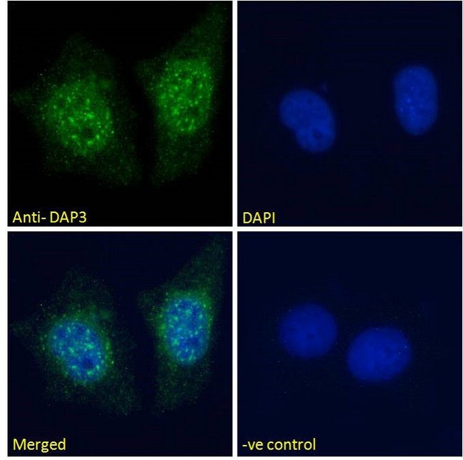
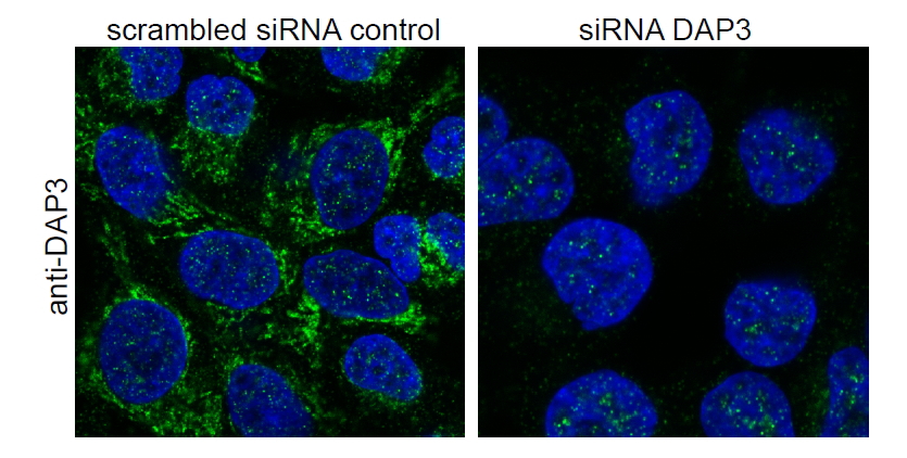
Validation
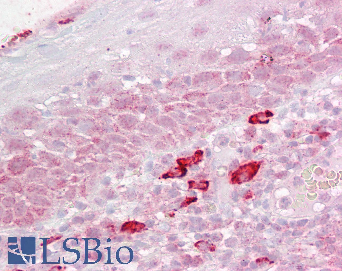
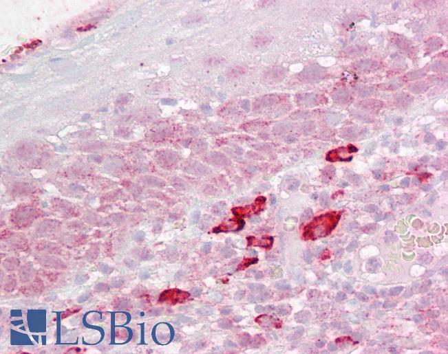
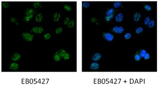
Anti-DAP3 antibody IHC of human tonsil. Immunohistochemistry of formalin-fixed, paraffin-embedded tissue after heat-induced antigen retrieval. Antibody concentration 2.5 ug/ml.
Anti-DAP3 antibody IHC of human tonsil. Immunohistochemistry of formalin-fixed, paraffin-embedded tissue after heat-induced antigen retrieval. Antibody concentration 2.5 ug/ml.
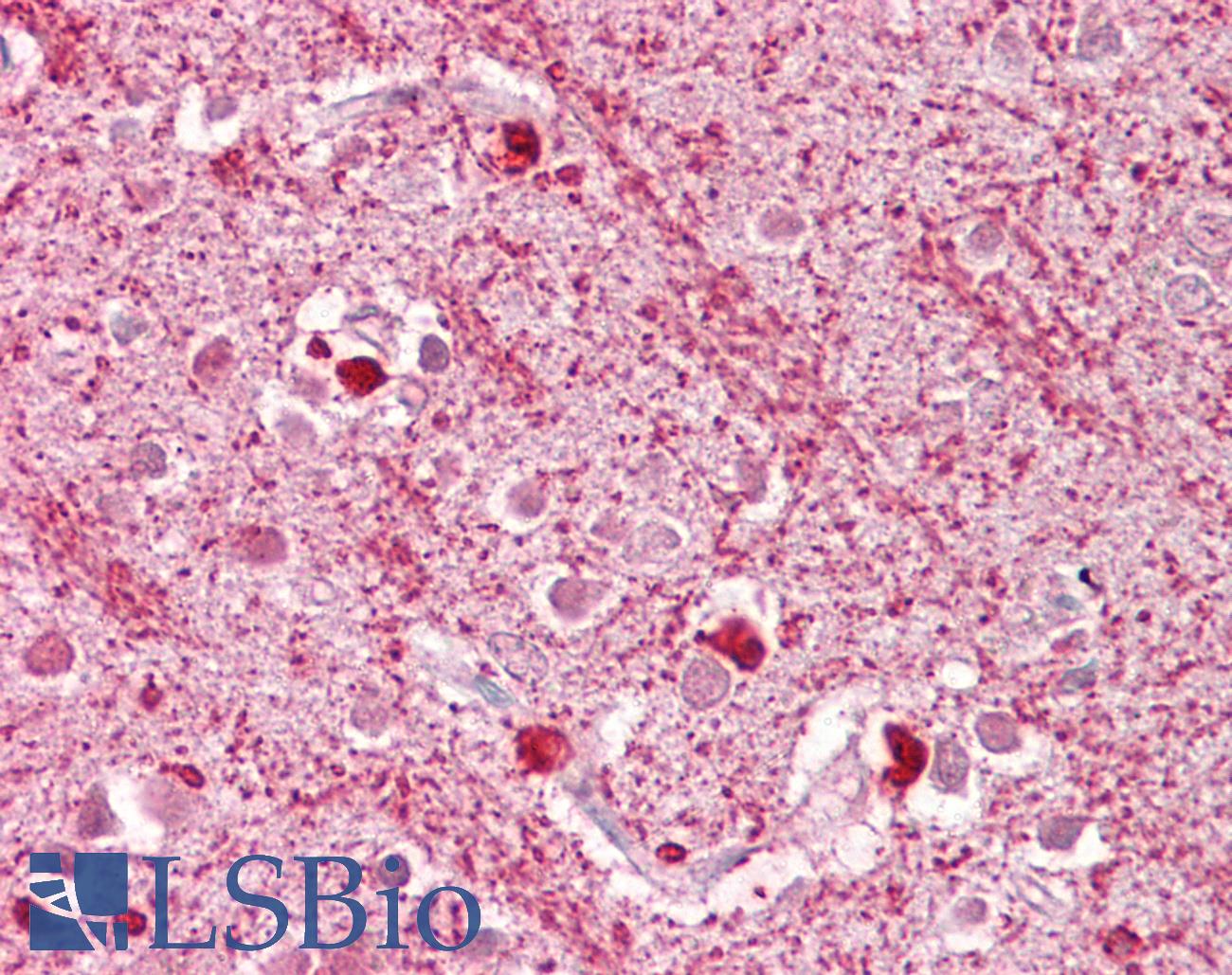
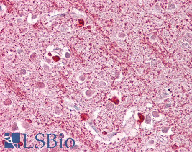
Anti-DAP3 antibody IHC of human brain, cortex. Immunohistochemistry of formalin-fixed, paraffin-embedded tissue after heat-induced antigen retrieval. Antibody concentration 2.5 ug/ml.
Anti-DAP3 antibody IHC of human brain, cortex. Immunohistochemistry of formalin-fixed, paraffin-embedded tissue after heat-induced antigen retrieval. Antibody concentration 2.5 ug/ml.
See More About...
LSBio Ratings
IHC-plus™ DAP3 Antibody (aa387-398) for IHC, IF/Immunofluorescence, WB/Western LS-B6446 has an LSBio Rating of
Laboratory Validation Score (4)
Learn more about The LSBio Ratings Algorithm
Publications (0)
Customer Reviews (0)
Featured Products


Reactivity:
All species
Range:
0.156-10 ng/ml
Reactivity:
Pig
Range:
78-5000 pg/ml
Source:
Hamster-Chinese
Tag:
His, N-Terminal
Request SDS/MSDS
To request an SDS/MSDS form for this product, please contact our Technical Support department at:
Technical.Support@LSBio.com
Requested From: United States
Date Requested: 4/18/2024
Date Requested: 4/18/2024


