Login
Registration enables users to use special features of this website, such as past
order histories, retained contact details for faster checkout, review submissions, and special promotions.
order histories, retained contact details for faster checkout, review submissions, and special promotions.
Forgot password?
Registration enables users to use special features of this website, such as past
order histories, retained contact details for faster checkout, review submissions, and special promotions.
order histories, retained contact details for faster checkout, review submissions, and special promotions.
Quick Order
Products
Antibodies
ELISA and Assay Kits
Research Areas
Infectious Disease
Resources
Purchasing
Reference Material
Contact Us
Locations
Orders Processing,
Shipping & Receiving,
Warehouse
2 Shaker Rd Suites
B001/B101
Shirley, MA 01464
Production Lab
Floor 6, Suite 620
20700 44th Avenue W
Lynnwood, WA 98036
Telephone Numbers
Tel: +1 (206) 374-1102
Fax: +1 (206) 577-4565
Contact Us
Additional Contact Details
Login
Registration enables users to use special features of this website, such as past
order histories, retained contact details for faster checkout, review submissions, and special promotions.
order histories, retained contact details for faster checkout, review submissions, and special promotions.
Forgot password?
Registration enables users to use special features of this website, such as past
order histories, retained contact details for faster checkout, review submissions, and special promotions.
order histories, retained contact details for faster checkout, review submissions, and special promotions.
Quick Order
| Catalog Number | Size | Price |
|---|---|---|
| LS-B342-50 | 50 µg (1 mg/ml) | $695 |
1 of 3
2 of 3
3 of 3
IHC‑plus™ Polyclonal Rabbit anti‑Mammal Collagen VI Antibody (IHC, WB) LS‑B342
IHC‑plus™ Polyclonal Rabbit anti‑Mammal Collagen VI Antibody (IHC, WB) LS‑B342

Note: This antibody replaces LS-C18865
Antibody:
Collagen VI Rabbit anti-Mammal Polyclonal Antibody
Application:
IHC, IHC-P, WB, IP, ELISA, FLISA
Reactivity:
Mammal, Human, Bovine
Format:
Unconjugated, Unmodified
Toll Free North America
 206-374-1102
For Research Use Only
Overview
Antibody:
Collagen VI Rabbit anti-Mammal Polyclonal Antibody
Application:
IHC, IHC-P, WB, IP, ELISA, FLISA
Reactivity:
Mammal, Human, Bovine
Format:
Unconjugated, Unmodified
Specifications
Description
Collagen VI antibody LS-B342 is an unconjugated rabbit polyclonal antibody to Collagen VI from mammal. It is reactive with human, bovine and mammal. Validated for ELISA, FLISA, IHC, IP and WB. Cited in 5 publications.
Host
Rabbit
Reactivity
Mammal, Human, Bovine
(tested or 100% immunogen sequence identity)
Clonality
IgG
Polyclonal
Conjugations
Unconjugated
Purification
Affinity chromatography
Modifications
Unmodified
Immunogen
Collagen Type I from human and bovine placenta.
Specificity
Typically negligible cross reactivity against other types of collagens was detected by ELISA against purified standards. Some class-specific anti-collagens may be specific for three-dimensional epitopes which may result in diminished reactivity with denatured collagen or formalin-fixed, paraffin embedded tissues. This antibody reacts with human, bovine, and most mammalian Type I collagens with negligible cross-reactivity with Type II, III, IV, V or VI collagens. Non-specific cross-reaction of anti-collagen antibodies with other human serum proteins or non-collagen extracellular matrix proteins is negligible.
Applications
- IHC
- IHC - Paraffin (2.5 µg/ml)
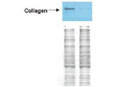
- Western blot (1:1000 - 1:10000)
- Immunoprecipitation (1:100)
- ELISA (1:5000 - 1:50000)
- Fluorophore-Linked Immunosorbent Assay (1:100)

|
Performing IHC? See our complete line of Immunohistochemistry Reagents including antigen retrieval solutions, blocking agents
ABC Detection Kits and polymers, biotinylated secondary antibodies, substrates and more.
|
Usage
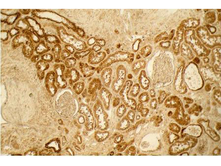
Immunohistochemistry: LS-B342 was validated for use in immunohistochemistry on a panel of 21 formalin-fixed, paraffin-embedded (FFPE) human tissues after heat induced antigen retrieval in pH 6.0 citrate buffer. After incubation with the primary antibody, slides were incubated with biotinylated secondary antibody, followed by alkaline phosphatase-streptavidin and chromogen. The stained slides were evaluated by a pathologist to confirm staining specificity. The optimal working concentration for LS-B342 was determined to be 2.5 ug/ml.
Presentation
0.02 M Potassium Phosphate, pH 7.2, 0.15 M NaCl, 0.01% Sodium Azide
Storage
Short term: store at 4°C. Long term: store at -20°C. Avoid freeze-thaw cycles.
Restrictions
For research use only. Intended for use by laboratory professionals.
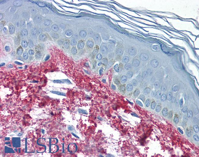
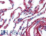
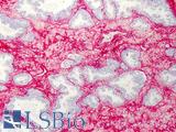
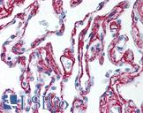
Validation

Anti-Collagen I antibody IHC of human skin. Immunohistochemistry of formalin-fixed, paraffin-embedded tissue after heat-induced antigen retrieval. Antibody concentration 5 ug/ml.
Anti-Collagen I antibody IHC of human skin. Immunohistochemistry of formalin-fixed, paraffin-embedded tissue after heat-induced antigen retrieval. Antibody concentration 5 ug/ml.
See More About...
LSBio Ratings
IHC-plus™ Collagen VI Antibody for IHC, WB/Western, IP, ELISA LS-B342 has an LSBio Rating of
Publications (4.4)
Laboratory Validation Score (4)
Learn more about The LSBio Ratings Algorithm
Publications (5)
Inhibition of collagen alpha 1(I) expression by the 5' stem-loop as a molecular decoy. Stefanovic B, Schnabl B, Brenner DA. The Journal of biological chemistry. 2002 277:18229-37.
Bone marrow-derived progenitor cells in pulmonary fibrosis. Hashimoto N, Jin H, Liu T, Chensue SW, Phan SH. The Journal of clinical investigation. 2004 113:243-52.
Adipogenic transcriptional regulation of hepatic stellate cells. She H, Xiong S, Hazra S, Tsukamoto H. The Journal of biological chemistry. 2005 280:4959-67.
Hepatic steatosis, fibrosis, and cancer in elderly cadavers. Mak KM, Kwong AJ, Chu E, Hoo NM. Anatomical record (Hoboken, N.J. : 2007). 2012 295:40-50.
Subtype-Specific Tumor-Associated Fibroblasts Contribute to the Pathogenesis of Uterine Leiomyoma. Wu X, Serna VA, Thomas J, Qiang W, Blumenfeld ML, Kurita T. Cancer research. 2017 December;77:6891-6901.
Customer Reviews (0)
Featured Products
Species:
Mammal, Human, Bovine
Applications:
IHC, IHC - Paraffin, Western blot, Immunoprecipitation, ELISA
Species:
Mammal, Human, Bovine
Applications:
IHC, IHC - Paraffin, Western blot, Immunoprecipitation, ELISA
Species:
Mammal, Human, Bovine
Applications:
IHC, IHC - Paraffin, Western blot, Immunoprecipitation, ELISA
Species:
Human, Rabbit
Applications:
IHC, IHC - Frozen, Western blot, ELISA
Species:
Human
Applications:
IHC, IHC - Frozen, Western blot, Immunoprecipitation, ELISA
Request SDS/MSDS
To request an SDS/MSDS form for this product, please contact our Technical Support department at:
Technical.Support@LSBio.com
Requested From: United States
Date Requested: 4/24/2024
Date Requested: 4/24/2024


