Login
Registration enables users to use special features of this website, such as past
order histories, retained contact details for faster checkout, review submissions, and special promotions.
order histories, retained contact details for faster checkout, review submissions, and special promotions.
Forgot password?
Registration enables users to use special features of this website, such as past
order histories, retained contact details for faster checkout, review submissions, and special promotions.
order histories, retained contact details for faster checkout, review submissions, and special promotions.
Quick Order
Products
Antibodies
ELISA and Assay Kits
Research Areas
Infectious Disease
Resources
Purchasing
Reference Material
Contact Us
Locations
Orders Processing,
Shipping & Receiving,
Warehouse
2 Shaker Rd Suites
B001/B101
Shirley, MA 01464
Production Lab
Floor 6, Suite 620
20700 44th Avenue W
Lynnwood, WA 98036
Telephone Numbers
Tel: +1 (206) 374-1102
Fax: +1 (206) 577-4565
Contact Us
Additional Contact Details
Login
Registration enables users to use special features of this website, such as past
order histories, retained contact details for faster checkout, review submissions, and special promotions.
order histories, retained contact details for faster checkout, review submissions, and special promotions.
Forgot password?
Registration enables users to use special features of this website, such as past
order histories, retained contact details for faster checkout, review submissions, and special promotions.
order histories, retained contact details for faster checkout, review submissions, and special promotions.
Quick Order
| Catalog Number | Size | Price |
|---|---|---|
| LS-C396086-50 | 50 µg | $294 |
| LS-C396086-100 | 100 µg | $360 |
1 of 5
2 of 5
3 of 5
4 of 5
5 of 5
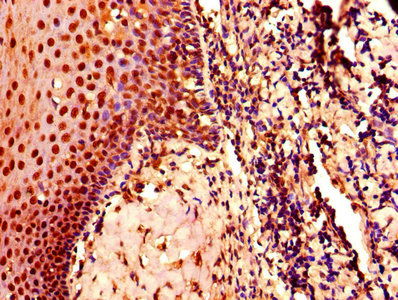
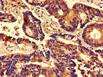
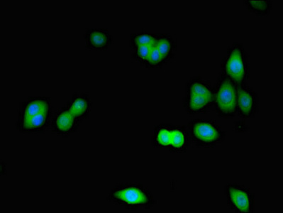
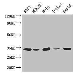
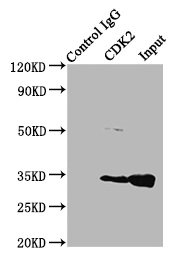
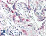
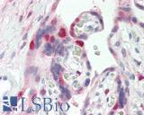
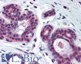
Polyclonal Rabbit anti‑Human CDK2 Antibody LS‑C396086
Polyclonal Rabbit anti‑Human CDK2 Antibody LS‑C396086
Toll Free North America
 206-374-1102
For Research Use Only
Overview
Specifications
Description
CDK2 antibody LS-C396086 is an unconjugated rabbit polyclonal antibody to human CDK2. Validated for ELISA.
Target
Human CDK2
Synonyms
CDK2 | Cell division protein kinase 2 | CDC2-related protein kinase | p33 protein kinase | p33(CDK2) | CDKN2 | Cyclin-dependent kinase 2
Host
Rabbit
Reactivity
Human
(tested or 100% immunogen sequence identity)
Clonality
IgG
Polyclonal
Purification
Protein G purified
Modifications
Unmodified
Immunogen
Recombinant human Cyclin-dependent kinase 2 protein.
Specificity
Human CDK2
Applications
- ELISA
Presentation
PBS, pH 7.4, 0.03% Proclin 300, 50% glycerol.
Storage
Short term: -20°C; Long term: -80°C; Avoid freeze-thaw cycles.
Restrictions
For research use only. Intended for use by laboratory professionals.
About CDK2
Publications (0)
Customer Reviews (0)
Featured Products
Species:
Human, Mouse, Xenopus
Applications:
IHC, IHC - Paraffin, Western blot, Immunoprecipitation, ELISA
Species:
Human, Mouse, Rat
Applications:
IHC, IHC - Paraffin, Immunofluorescence, Western blot, Immunoprecipitation, ELISA
Species:
Human, Mouse, Rat
Applications:
IHC, IHC - Paraffin, Immunofluorescence, Western blot, Immunoprecipitation, ELISA
Species:
Human, Mouse
Applications:
IHC, IHC - Paraffin, Western blot, Immunoprecipitation
Species:
Human
Applications:
Western blot, Peptide Enzyme-Linked Immunosorbent Assay
Request SDS/MSDS
To request an SDS/MSDS form for this product, please contact our Technical Support department at:
Technical.Support@LSBio.com
Requested From: United States
Date Requested: 4/24/2024
Date Requested: 4/24/2024


