order histories, retained contact details for faster checkout, review submissions, and special promotions.
Forgot password?
order histories, retained contact details for faster checkout, review submissions, and special promotions.
Locations
Orders Processing,
Shipping & Receiving,
Warehouse
2 Shaker Rd Suites
B001/B101
Shirley, MA 01464
Production Lab
Floor 6, Suite 620
20700 44th Avenue W
Lynnwood, WA 98036
Telephone Numbers
Tel: +1 (206) 374-1102
Fax: +1 (206) 577-4565
Contact Us
Additional Contact Details
order histories, retained contact details for faster checkout, review submissions, and special promotions.
Forgot password?
order histories, retained contact details for faster checkout, review submissions, and special promotions.
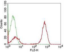
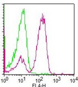
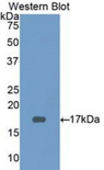
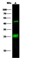
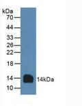
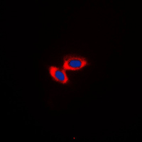
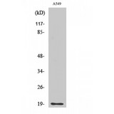
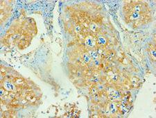
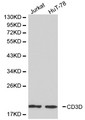
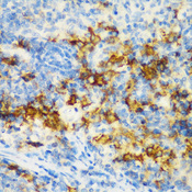
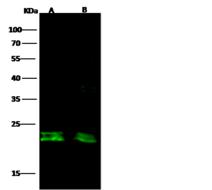
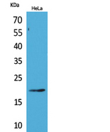
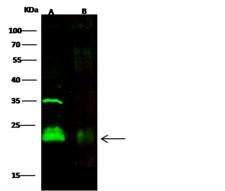
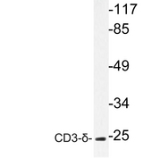
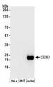
CD3D
CD3d molecule, delta (CD3-TCR complex)
CD3D is part of the T-cell receptor/CD3 complex (TCR/CD3 complex) and is involved in T-cell development and signal transduction. The encoded membrane protein represents the delta subunit of the CD3 complex, and along with four other CD3 subunits, binds either TCR alpha/beta or TCR gamma/delta to form the TCR/CD3 complex on the surface of T-cells. Defects in this gene are a cause of severe combined immunodeficiency autosomal recessive T-cell-negative/B-cell-positive/NK-cell-positive (SCIDBNK). Two transcript variants encoding different isoforms have been found for this gene. Other variants may also exist, but the full-length natures of their transcripts has yet to be defined.
| Gene Name: | CD3d molecule, delta (CD3-TCR complex) |
| Synonyms: | CD3D, CD3 delta, CD3d antigen, T-cell receptor T3 delta chain, T3D, CD3 antigen, delta subunit, CD3-DELTA, OKT3, delta chain |
| Target Sequences: | NM_000732 NP_000723.1 P04234 |

If you do not find the reagent or information you require, please contact Customer.Support@LSBio.com to inquire about additional products in development.


