Login
Registration enables users to use special features of this website, such as past
order histories, retained contact details for faster checkout, review submissions, and special promotions.
order histories, retained contact details for faster checkout, review submissions, and special promotions.
Forgot password?
Registration enables users to use special features of this website, such as past
order histories, retained contact details for faster checkout, review submissions, and special promotions.
order histories, retained contact details for faster checkout, review submissions, and special promotions.
Quick Order
Products
Antibodies
ELISA and Assay Kits
Research Areas
Infectious Disease
Resources
Purchasing
Reference Material
Contact Us
Locations
Orders Processing,
Shipping & Receiving,
Warehouse
2 Shaker Rd Suites
B001/B101
Shirley, MA 01464
Production Lab
Floor 6, Suite 620
20700 44th Avenue W
Lynnwood, WA 98036
Telephone Numbers
Tel: +1 (206) 374-1102
Fax: +1 (206) 577-4565
Contact Us
Additional Contact Details
Login
Registration enables users to use special features of this website, such as past
order histories, retained contact details for faster checkout, review submissions, and special promotions.
order histories, retained contact details for faster checkout, review submissions, and special promotions.
Forgot password?
Registration enables users to use special features of this website, such as past
order histories, retained contact details for faster checkout, review submissions, and special promotions.
order histories, retained contact details for faster checkout, review submissions, and special promotions.
Quick Order
| Catalog Number | Size | Price |
|---|---|---|
| LS-C209224-100 | 100 µg (1 mg/ml) | $544 |
1 of 2
2 of 2
Polyclonal Rabbit anti‑E. coli Beta Galactosidase Antibody (IHC, IF, WB) LS‑C209224
Polyclonal Rabbit anti‑E. coli Beta Galactosidase Antibody (IHC, IF, WB) LS‑C209224
Antibody:
Beta Galactosidase Rabbit anti-E. coli Polyclonal Antibody
Application:
IHC, IF, WB, IP, ELISA
Reactivity:
E. coli
Format:
Unconjugated, Unmodified
Toll Free North America
 206-374-1102
For Research Use Only
Overview
Antibody:
Beta Galactosidase Rabbit anti-E. coli Polyclonal Antibody
Application:
IHC, IF, WB, IP, ELISA
Reactivity:
E. coli
Format:
Unconjugated, Unmodified
Specifications
Description
Beta Galactosidase antibody LS-C209224 is an unconjugated rabbit polyclonal antibody to e. coli Beta Galactosidase. Validated for ELISA, IF, IHC, IP and WB.
Host
Rabbit
Reactivity
E. coli
(tested or 100% immunogen sequence identity)
Clonality
IgG
Polyclonal
Conjugations
Unconjugated
Purification
Delipidation, salt fractionation and ion exchange chromatography followed by dialysis.
Modifications
Unmodified
Immunogen
Full length native Beta Galactosidase isolated from E.coli
Specificity
Assay by immunoelectrophoresis resulted in a single precipitin arc against anti-Rabbit Serum as well as purified and partially purified Beta Galactosidase [E.coli]. Cross reactivity against Beta Galactosidase from other tissues and species may occur but have not been specifically determined. Very low background staining has been reported in various assays.
Applications
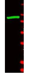
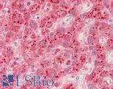
- IHC (1:1500)
- Immunofluorescence
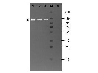
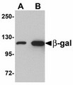
- Western blot (1:5000 - 1:10000)
- Immunoprecipitation
- ELISA (1:10000)

|
Performing IHC? See our complete line of Immunohistochemistry Reagents including antigen retrieval solutions, blocking agents
ABC Detection Kits and polymers, biotinylated secondary antibodies, substrates and more.
|
Usage
Beta-Gal Antibody is suitable for immunoblotting (western or dot blot), ELISA, immunofluorescence microscopy, immunoprecipitation, conjugation and most immunological methods requiring high titer and specificity. The antibody recognizes both frozen tissue sections, paraffin embedded tissue and 4% paraformaldehyde fixed tissue for most immunohistochemical analysis. A 1:1 dilution has been reported to detect beta-galactosidase in adult rat spinal cord tissue after infection with helper-dependent adenovirus expressing lacZ. In this particular experiment, tissue was perfused with 4% paraformaldehyde and cryostat-cut (35 mu m) to produce free-floating sections.
Presentation
Lyophilized from 0.02 M Potassium Phosphate, pH 7.2, 0.15 M NaCl, 0.01% Sodium Azide
Reconstitution
Reconstitute in 100 µL of deionized water.
Storage
Short term 4°C, long term aliquot and store at -20°C, avoid freeze-thaw cycles. Centrifuge product before removing cap. Only dilute immediately prior to use.
Restrictions
For research use only. Intended for use by laboratory professionals.
Publications (0)
Customer Reviews (0)
Featured Products
Species:
E. coli
Applications:
ICC, Immunofluorescence, Western blot, ELISA
Species:
Human
Applications:
IHC, IHC - Paraffin, Western blot
Species:
E. coli
Applications:
Immunofluorescence, Western blot, ELISA
Request SDS/MSDS
To request an SDS/MSDS form for this product, please contact our Technical Support department at:
Technical.Support@LSBio.com
Requested From: United States
Date Requested: 4/25/2024
Date Requested: 4/25/2024


