Login
Registration enables users to use special features of this website, such as past
order histories, retained contact details for faster checkout, review submissions, and special promotions.
order histories, retained contact details for faster checkout, review submissions, and special promotions.
Forgot password?
Registration enables users to use special features of this website, such as past
order histories, retained contact details for faster checkout, review submissions, and special promotions.
order histories, retained contact details for faster checkout, review submissions, and special promotions.
Quick Order
Products
Antibodies
ELISA and Assay Kits
Research Areas
Infectious Disease
Resources
Purchasing
Reference Material
Contact Us
Locations
Orders Processing,
Shipping & Receiving,
Warehouse
2 Shaker Rd Suites
B001/B101
Shirley, MA 01464
Production Lab
Floor 6, Suite 620
20700 44th Avenue W
Lynnwood, WA 98036
Telephone Numbers
Tel: +1 (206) 374-1102
Fax: +1 (206) 577-4565
Contact Us
Additional Contact Details
Login
Registration enables users to use special features of this website, such as past
order histories, retained contact details for faster checkout, review submissions, and special promotions.
order histories, retained contact details for faster checkout, review submissions, and special promotions.
Forgot password?
Registration enables users to use special features of this website, such as past
order histories, retained contact details for faster checkout, review submissions, and special promotions.
order histories, retained contact details for faster checkout, review submissions, and special promotions.
Quick Order
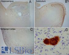
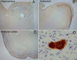
Amyloid Fibrils
Amyloid Fibrils Target Details
| Target Name: | Amyloid Fibrils |
☰ Filters
Products
Antibodies
(12)
Type
Primary
(12)
Target
Amyloid Fibrils
(12)
Reactivity
Human
(12)
Mouse
(2)
Rat
(2)
Application
IHC
(12)
WB
(12)
DB
(12)
ELISA
(12)
ICC
(10)
IP
(10)
Host
rabbit
(12)
Clonality
polyclonal pc
(12)
Format
AP Conjugated
(1)
APC Conjugated
(1)
Atto 390 Conjugated
(1)
Atto 488 Conjugated
(1)
Atto 594 Conjugated
(1)
Biotin Conjugated
(1)
FITC Conjugated
(1)
PerCP Conjugated
(1)
RPE Conjugated
(1)
Unconjugated
(3)
Publications
No
(12)

Amyloid Fibrils Rabbit Polyclonal Antibody
Mouse, Rat, Human
DB, ELISA, ICC, IHC, IP, WB
Unconjugated
100 µl/$433

Amyloid Fibrils Rabbit anti-Human Polyclonal (Biotin) Antibody
Human
DB, ELISA, ICC, IHC, IP, WB
Biotin Conjugated
100 µl/$467

Amyloid Fibrils Rabbit anti-Human Polyclonal (FITC) Antibody
Human
DB, ELISA, ICC, IHC, IP, WB
FITC Conjugated
100 µl/$463

Amyloid Fibrils Rabbit anti-Human Polyclonal (PerCP) Antibody
Human
DB, ELISA, ICC, IHC, IP, WB
PerCP Conjugated
100 µl/$470

Amyloid Fibrils Rabbit anti-Human Polyclonal (RPE) Antibody
Human
DB, ELISA, ICC, IHC, IP, WB
RPE Conjugated
100 µl/$467

Amyloid Fibrils Rabbit anti-Human Polyclonal (Atto 488) Antibody
Human
DB, ELISA, ICC, IHC, IP, WB
Atto 488 Conjugated
100 µl/$470

Amyloid Fibrils Rabbit anti-Human Polyclonal (Atto 390) Antibody
Human
DB, ELISA, ICC, IHC, IP, WB
Atto 390 Conjugated
100 µl/$471
Amyloid Fibrils Rabbit anti-Human Polyclonal (Atto 594) Antibody
Human
DB, ELISA, ICC, IHC, IP, WB
Atto 594 Conjugated
100 µl/$470

Amyloid Fibrils Rabbit anti-Human Polyclonal (APC) Antibody
Human
DB, ELISA, ICC, IHC, IP, WB
APC Conjugated
100 µl/$470

Amyloid Fibrils Rabbit anti-Human Polyclonal (AP) Antibody
Human
DB, ELISA, ICC, IHC, IP, WB
AP Conjugated
100 µl/$465
Amyloid Fibrils Rabbit anti-Human Polyclonal Antibody
Human
DB, ELISA, IHC, WB
Unconjugated
50 µl/$569
Amyloid Fibrils Rabbit anti-Human Polyclonal Antibody
Mouse, Rat, Human
DB, ELISA, IHC, WB
Unconjugated
100 µl/$531
Viewing 1-12
of 12
product results
If you do not find the reagent or information you require, please contact Customer.Support@LSBio.com to inquire about additional products in development.


