Login
Registration enables users to use special features of this website, such as past
order histories, retained contact details for faster checkout, review submissions, and special promotions.
order histories, retained contact details for faster checkout, review submissions, and special promotions.
Forgot password?
Registration enables users to use special features of this website, such as past
order histories, retained contact details for faster checkout, review submissions, and special promotions.
order histories, retained contact details for faster checkout, review submissions, and special promotions.
Quick Order
Products
Antibodies
ELISA and Assay Kits
Research Areas
Infectious Disease
Resources
Purchasing
Reference Material
Contact Us
Locations
Orders Processing,
Shipping & Receiving,
Warehouse
2 Shaker Rd Suites
B001/B101
Shirley, MA 01464
Production Lab
Floor 6, Suite 620
20700 44th Avenue W
Lynnwood, WA 98036
Telephone Numbers
Tel: +1 (206) 374-1102
Fax: +1 (206) 577-4565
Contact Us
Additional Contact Details
Login
Registration enables users to use special features of this website, such as past
order histories, retained contact details for faster checkout, review submissions, and special promotions.
order histories, retained contact details for faster checkout, review submissions, and special promotions.
Forgot password?
Registration enables users to use special features of this website, such as past
order histories, retained contact details for faster checkout, review submissions, and special promotions.
order histories, retained contact details for faster checkout, review submissions, and special promotions.
Quick Order
| Catalog Number | Size | Price |
|---|---|---|
| LS-C775549-100 | 100 µg (1 mg/ml) | $497 |
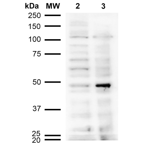
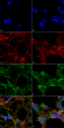
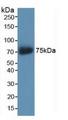
![Acrolein Antibody - Mouse Anti-Acrolein Antibody [10A10] used in Western Blot (WB) on Cervical cancer cell line (HeLa) lysate](https://lsbio-7d62.kxcdn.com/image2/acrolein-antibody-clone-10a10-ls-c775549/514525_4193267.png)
1 of 5
2 of 5
3 of 5
4 of 5
5 of 5
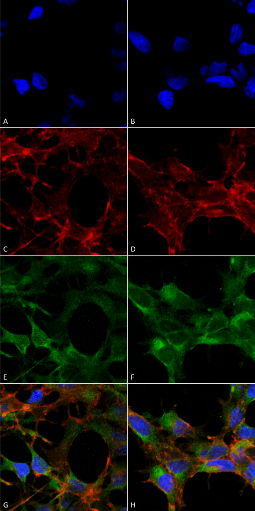
Monoclonal Mouse anti‑All species Acrolein Antibody (clone 10A10, IF, WB) LS‑C775549
Monoclonal Mouse anti‑All species Acrolein Antibody (clone 10A10, IF, WB) LS‑C775549
Antibody:
Acrolein Mouse Monoclonal (10A10) Antibody
Application:
IF, WB, Flo, ELISA
Reactivity:
All species
Format:
Unconjugated, Unmodified
Toll Free North America
 206-374-1102
For Research Use Only
Overview
Antibody:
Acrolein Mouse Monoclonal (10A10) Antibody
Application:
IF, WB, Flo, ELISA
Reactivity:
All species
Format:
Unconjugated, Unmodified
Specifications
Description
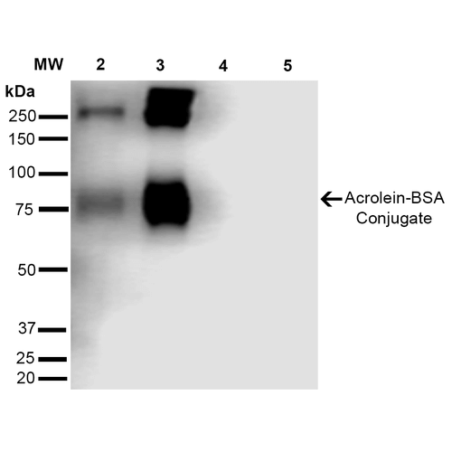
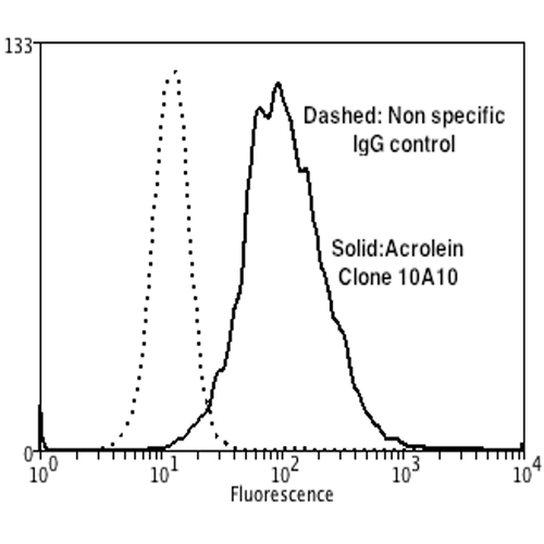
Acrolein antibody LS-C775549 is an unconjugated mouse monoclonal antibody to all species Acrolein. Validated for ELISA, Flow, IF and WB.
Host
Mouse
Reactivity
All species
Clonality
IgG1
Monoclonal
Clone
10A10
Conjugations
Unconjugated
Purification
Protein G purified
Modifications
Unmodified
Immunogen
Synthetic Acrolein modified Keyhole Limpet Hemocyanin (KLH).
Specificity
Specific for Acrolein modified proteins. Does not detect free acrolein. Does not cross-react with Crotonaldehyde, Hexanoyl Lysine, 4-Hydroxy-2-hexenal, 4-Hydroxy nonenal, Malondialdehyde, or Methylglyoxal modified proteins.
Applications
- Immunofluorescence (1:50)
- Western blot (1:1000)
- Flow Cytometry (1:50)
- ELISA (1:1000)
Usage
Applications should be user optimized.
Presentation
PBS, pH 7.4, 0.09% Sodium Azide, 50% Glycerol
Storage
Store at -20°C.
Restrictions
For research use only. Intended for use by laboratory professionals.
Publications (0)
Customer Reviews (0)
Featured Products
Species:
All species
Applications:
Immunofluorescence, Western blot, Flow Cytometry, ELISA
Species:
Human
Applications:
IHC, IHC - Paraffin, Western blot, ELISA
Reactivity:
Human
Range:
15.6-1000 pg/ml
Reactivity:
Monkey
Range:
0.156-10 ng/ml
Reactivity:
Human
Range:
0.156-10 ng/ml
Request SDS/MSDS
To request an SDS/MSDS form for this product, please contact our Technical Support department at:
Technical.Support@LSBio.com
Requested From: United States
Date Requested: 4/17/2024
Date Requested: 4/17/2024


