Login
Registration enables users to use special features of this website, such as past
order histories, retained contact details for faster checkout, review submissions, and special promotions.
order histories, retained contact details for faster checkout, review submissions, and special promotions.
Forgot password?
Registration enables users to use special features of this website, such as past
order histories, retained contact details for faster checkout, review submissions, and special promotions.
order histories, retained contact details for faster checkout, review submissions, and special promotions.
Quick Order
Products
Antibodies
ELISA and Assay Kits
Research Areas
Infectious Disease
Resources
Purchasing
Reference Material
Contact Us
Locations
Orders Processing,
Shipping & Receiving,
Warehouse
2 Shaker Rd Suites
B001/B101
Shirley, MA 01464
Production Lab
Floor 6, Suite 620
20700 44th Avenue W
Lynnwood, WA 98036
Telephone Numbers
Tel: +1 (206) 374-1102
Fax: +1 (206) 577-4565
Contact Us
Additional Contact Details
Login
Registration enables users to use special features of this website, such as past
order histories, retained contact details for faster checkout, review submissions, and special promotions.
order histories, retained contact details for faster checkout, review submissions, and special promotions.
Forgot password?
Registration enables users to use special features of this website, such as past
order histories, retained contact details for faster checkout, review submissions, and special promotions.
order histories, retained contact details for faster checkout, review submissions, and special promotions.
Quick Order
| Catalog Number | Size | Price |
|---|---|---|
| LS-C156406-400 | 400 µl | $393 |
1 of 5
2 of 5
3 of 5
4 of 5
5 of 5
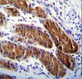
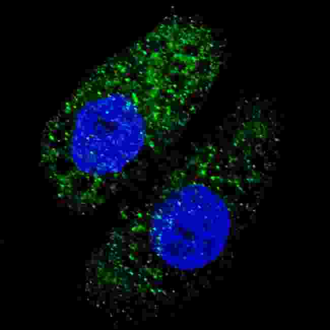
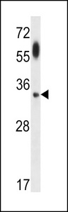
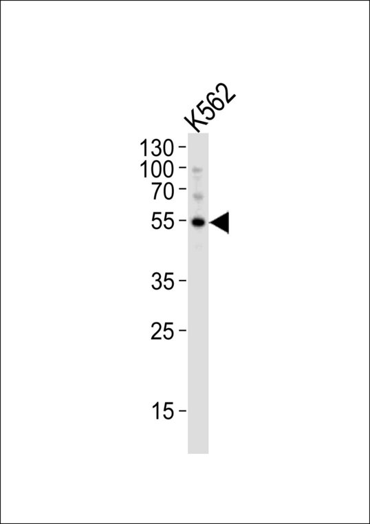
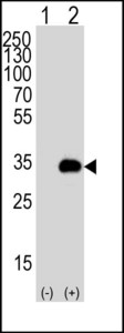
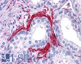
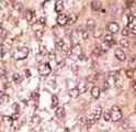
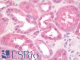
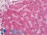
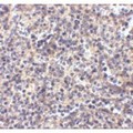
Polyclonal Rabbit anti‑Human APG5 / ATG5 Antibody (IHC, IF, WB) LS‑C156406
Polyclonal Rabbit anti‑Human APG5 / ATG5 Antibody (IHC, IF, WB) LS‑C156406

Antibody:
APG5 / ATG5 Rabbit anti-Human Polyclonal Antibody
Application:
IHC, IHC-P, IF, WB
Reactivity:
Human
Format:
Unconjugated, Unmodified
Toll Free North America
 206-374-1102
For Research Use Only
Overview
Antibody:
APG5 / ATG5 Rabbit anti-Human Polyclonal Antibody
Application:
IHC, IHC-P, IF, WB
Reactivity:
Human
Format:
Unconjugated, Unmodified
Specifications
Description
ATG5 antibody LS-C156406 is an unconjugated rabbit polyclonal antibody to human ATG5 (APG5). Validated for IF, IHC and WB. Cited in 1 publication.
Target
Human APG5 / ATG5
Synonyms
ATG5 | APG5 | APG5-LIKE | APG5L | Apoptosis specific protein | Apoptosis-specific protein | Autophagy related 5 | HAPG5 | ASP | Autophagy protein 5
Host
Rabbit
Reactivity
Human
(tested or 100% immunogen sequence identity)
Clonality
Polyclonal
Conjugations
Unconjugated
Purification
Protein A purified
Modifications
Unmodified
Applications
- IHC
- IHC - Paraffin (1:10 - 1:50)
- Immunofluorescence (1:200)
- Western blot (1:1000)

|
Performing IHC? See our complete line of Immunohistochemistry Reagents including antigen retrieval solutions, blocking agents
ABC Detection Kits and polymers, biotinylated secondary antibodies, substrates and more.
|
Presentation
PBS, 0.09% Sodium Azide
Storage
Maintain refrigerated at 2°C to 8°C for up to 6 months. For long term storage store at -20°C.
Restrictions
For research use only. Intended for use by laboratory professionals.
About APG5 / ATG5
LSBio Ratings
APG5 / ATG5 Antibody for IHC, IF/Immunofluorescence, WB/Western LS-C156406 has an LSBio Rating of
Publications (4)
Learn more about The LSBio Ratings Algorithm
Publications (1)
Autophagy induced by deficiency of sphingosine-1-phosphate phosphohydrolase 1 is switched to apoptosis by calpain-mediated autophagy-related gene 5 (Atg5) cleavage. Lpine S, Allegood JC, Edmonds Y, Milstien S, Spiegel S. The Journal of biological chemistry. 2011 286:44380-90.
Customer Reviews (0)
Featured Products
Species:
Human, Mouse, Rat, Bovine, Pig, Pufferfish, Zebrafish
Applications:
IHC, IHC - Paraffin
Species:
Human
Applications:
IHC, IHC - Paraffin, Western blot
Species:
Human, Mouse
Applications:
IHC, IHC - Paraffin, Immunofluorescence, Western blot
Species:
Human, Mouse
Applications:
IHC, IHC - Paraffin, Immunofluorescence, Western blot
Species:
Human, Mouse, Rat, Bovine, Pig, Xenopus, Zebrafish, Primate
Applications:
IHC, IHC - Paraffin, ICC, Western blot, Immunoprecipitation
Request SDS/MSDS
To request an SDS/MSDS form for this product, please contact our Technical Support department at:
Technical.Support@LSBio.com
Requested From: United States
Date Requested: 4/24/2024
Date Requested: 4/24/2024


